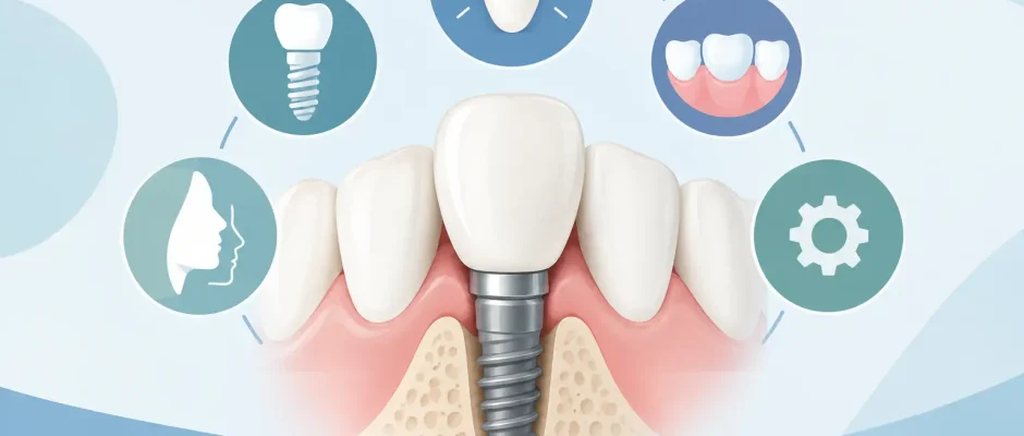
What Shapes a Front Tooth Implant Case Result?
A front tooth implant case is judged by more than whether the implant integrates with bone. When you speak, smile, or laugh, the eye notices the position of the tooth, the gum line, the tiny spaces between teeth, and how light passes through the crown. Replacing a front tooth is therefore both a surgical and an esthetic task. A stable implant is essential, but it is only the foundation for a result that should look calm, natural, and proportionate.
The best treatment plan begins before a tooth is removed. The goal is to preserve the tissues that frame the future crown, choose the right timing for implant placement, and avoid decisions that create compromises later.
Why a Front Tooth Implant Case Is Different
In the back of the mouth, implants must primarily withstand chewing forces and restore function. In the smile zone, function still matters, but the margin for visual error is much smaller. Even a one-millimeter difference in gum level or crown position may be noticeable.
The bone on the facial side of upper front teeth is often thin. After extraction, this bone naturally remodels and can shrink, especially if infection, trauma, or a long-standing missing tooth has already damaged the site. As the bone changes, the gum can recede. This may expose a gray shadow from the implant or create an uneven gum line compared with the neighboring tooth.
The papillae – the small triangular gum areas between teeth – are equally important. They affect whether dark spaces appear beside the crown. Their preservation depends on bone support around the adjacent natural teeth, soft-tissue thickness, implant position, and the shape of the temporary and final crowns.
For these reasons, a front tooth implant is not a procedure that should be planned from a two-dimensional X-ray alone. The surgeon must evaluate bone volume, root positions, bite, gum phenotype, smile line, and the condition of the neighboring teeth.
Diagnosis Comes Before the Surgical Date
A careful consultation usually includes a clinical examination, photographs, digital scans, and three-dimensional imaging when indicated. A CBCT scan helps assess the socket, the thickness of the facial bone wall, possible inflammation, and the amount of bone available beyond the root tip. It also helps determine whether an implant can be placed in a prosthetically correct position rather than simply where bone happens to be available.
The ideal implant position is dictated by the future crown. If an implant is placed too far toward the lip, the facial bone and gum may become unstable. If it is placed too far toward the palate, the crown may need an unnatural shape to meet the bite. If it is too close to an adjacent root, it can compromise the bone between teeth and affect the papilla.
Digital planning and a surgical guide can improve control over depth, angle, and three-dimensional position. A guide is not a substitute for surgical judgment, particularly in a fresh extraction socket where anatomy can change during the procedure. It is a tool that supports a precise, preplanned protocol.
Immediate Implant or a Staged Approach?
Patients often ask whether an implant can be placed on the same day as extraction. In selected cases, immediate implant placement can shorten the treatment timeline and help preserve the contours of the socket. It may be considered when the infection is manageable, the socket walls are sufficiently intact, primary implant stability can be achieved, and the gum tissue is favorable.
However, immediate placement is not automatically the best choice. A tooth with a large facial bone defect, active acute infection, severe gum recession, or inadequate bone for stable fixation may require a staged approach. In that situation, extraction is performed as gently as possible, often with socket preservation or bone grafting. Implant placement follows after healing, once the foundation is more predictable.
Choosing a staged protocol is not a setback. It can be the safer route to a better long-term esthetic result. The right question is not, “Can an implant be placed today?” but, “Which sequence gives this specific site the best chance of stable bone and natural gum contours?”
Protecting Bone and Soft Tissue During Surgery
Atraumatic extraction is a central part of treatment when a front tooth cannot be saved. The aim is to remove the root while preserving the thin socket walls and surrounding gums. This requires controlled technique and, in difficult cases, careful sectioning of the tooth rather than forceful elevation.
When an implant is placed immediately, a small gap commonly remains between the implant and the facial socket wall. This space may be filled with a bone substitute to support contour preservation. Depending on the anatomy, a membrane, connective-tissue graft, or platelet-rich fibrin (PRF) may also be used. PRF is produced from the patient’s own blood and can support soft-tissue healing as part of a broader regenerative protocol.
Soft-tissue grafting is not necessary in every case, but it is particularly valuable when the gum is thin, recession is present, or the smile line is high. Thick, well-supported tissue is generally more resistant to future recession and better able to mask underlying implant components.
The Temporary Crown Has a Clinical Role
A temporary tooth is not only about appearance while the site heals. In many front tooth cases, it helps guide the shape of the gums around the future crown. This is known as creating an emergence profile: the gradual transition from implant to tooth-shaped crown.
A temporary crown may be supported by the implant, bonded to neighboring teeth, or incorporated into a removable appliance. The correct option depends on implant stability, bite forces, grafting needs, and the patient’s habits. If the implant receives an immediate temporary crown, it is usually kept out of direct biting contacts during early healing. It should protect the tissues, not overload the implant.
The temporary is adjusted gradually as healing progresses. Small changes can encourage the gums to develop a more natural contour. Rushing this stage can lead to pressure on delicate tissues, recession, or an artificial-looking crown shape.
Selecting the Final Crown
The final restoration is typically planned after the implant has integrated and the gums have matured. The material and construction depend on the case. In the esthetic zone, ceramic restorations are often chosen for their ability to reflect light similarly to natural enamel. The abutment – the component connecting implant and crown – may also be selected to avoid a dark appearance through thin gum tissue.
Color matching deserves time and attention. A natural front tooth is rarely one uniform shade. It may have translucency near the edge, subtle surface texture, and small variations in color. The dental laboratory needs accurate photographs, shade information, and digital or conventional impressions to reproduce these details.
The final crown should also be designed for function. A beautiful crown that receives excessive bite forces can chip, loosen, or place stress on the implant and surrounding bone. Bite adjustment is especially relevant for patients who clench or grind their teeth. In some cases, a night guard is recommended after treatment.
What Can Affect Long-Term Stability?
Even well-executed surgery cannot remove every biological risk. Smoking, uncontrolled diabetes, untreated gum disease, poor plaque control, and heavy grinding can affect healing and long-term implant health. A history of periodontal disease does not necessarily rule out implant treatment, but inflammation must be controlled before treatment and monitored afterward.
Regular professional maintenance is part of protecting the result. Implant tissues can develop inflammation, just as natural teeth can develop gum disease. Patients should attend follow-up visits, clean carefully around the crown, and report bleeding, swelling, a change in bite, or crown mobility without delay.
The neighboring teeth also matter. If a nearby tooth has gum recession, an unstable old restoration, or periodontal bone loss, it can influence the esthetic outcome around the implant. Sometimes treating the surrounding teeth first or coordinating care with a restorative dentist produces a more balanced result.
A Predictable Result Is Built in Stages
A successful front tooth restoration depends on a coordinated sequence: diagnosis, careful extraction when needed, three-dimensional implant placement, management of bone and soft tissue, a properly designed temporary, and a final crown created around stable gums. The process may take several months, particularly when regeneration is required. That time is often what allows the tissues to heal without being forced into an unrealistic schedule.
For patients considering treatment in Tel Aviv, the consultation should end with a clear plan: what can be done immediately, what should heal first, which materials or grafting procedures may be needed, and what the expected stages will be. A clear explanation reduces uncertainty and helps patients make decisions based on biology rather than pressure.
A front tooth can be replaced beautifully, but the most natural results begin with restraint: preserving what can be preserved, rebuilding what has been lost, and allowing healing to guide the final design.


