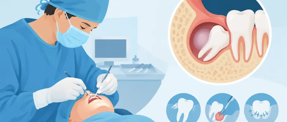
What Does Wisdom Tooth Cyst Surgery Involve?
A cyst discovered near a wisdom tooth is not simply a more complicated tooth extraction. It is a finding inside the jawbone that needs a precise diagnosis, a surgical plan, and follow-up based on healing and pathology. Wisdom tooth cyst surgery is performed to remove the cystic tissue, address the involved tooth when indicated, and reduce the risk of continued bone loss, infection, damage to adjacent teeth, or recurrence.
For many patients, the cyst is found unexpectedly on a panoramic X-ray taken before wisdom tooth removal. Often there is little or no pain. That absence of symptoms can be misleading: some jaw cysts expand slowly and quietly, thinning the bone long before they cause swelling, pressure, or infection.
When a cyst near a wisdom tooth needs treatment
The most common cyst associated with an unerupted wisdom tooth is a dentigerous cyst. It develops around the crown of a tooth that has remained within the jawbone, usually a lower third molar. On an X-ray, it may look like a dark, well-defined space surrounding the tooth. But imaging alone cannot always establish the final diagnosis.
Other lesions can resemble a dentigerous cyst, including an odontogenic keratocyst, inflammatory cyst, or, less commonly, another type of jaw lesion. This is why the tissue removed during surgery is typically sent for histopathologic examination. The laboratory result confirms what was treated and helps determine whether additional monitoring is needed.
Treatment is generally recommended when the lesion is enlarging, associated with pain or recurrent infection, affecting the roots of neighboring teeth, weakening the jawbone, or lying close to critical structures. Even a small, symptom-free lesion may warrant treatment if its appearance and location suggest a risk of progression.
Planning wisdom tooth cyst surgery safely
A panoramic X-ray is useful for initial assessment, but three-dimensional imaging is often the key step in surgical planning. A CBCT scan shows the true size of the cavity, the position of the wisdom tooth, the thickness of surrounding bone, and its relationship to the inferior alveolar nerve in the lower jaw or the maxillary sinus in the upper jaw.
This information changes the surgical approach. A cyst around a lower wisdom tooth may sit very close to the nerve responsible for sensation in the lower lip and chin. In the upper jaw, the surgical team evaluates the sinus floor and the risk of communication with the sinus. Careful planning does not eliminate every risk, but it allows the surgeon to choose the least traumatic and most predictable route.
The consultation should also include a review of medications, smoking or vaping, diabetes control, bleeding history, allergies, and previous jaw surgery. These factors affect anesthesia, infection risk, and the pace of healing. A clear plan is more reassuring than vague promises: patients should know what will be removed, whether a bone graft may be needed, how the specimen will be examined, and what follow-up is expected.
Will the wisdom tooth always be removed?
In most cases, the involved wisdom tooth is removed at the same time as the cyst. Leaving a tooth that is the source or center of the lesion can increase the chance of persistent disease or recurrence.
There are exceptions. If a deeply positioned lower wisdom tooth is intimately attached to the mandibular nerve, complete removal may carry a meaningful risk of sensory disturbance. In selected cases, a surgeon may discuss alternatives such as staged treatment, decompression of a large cyst, or coronectomy, where the crown is removed while roots close to the nerve are retained. The correct choice depends on the diagnosis, anatomy, symptoms, and long-term risk – not on a one-size-fits-all protocol.
What happens during the procedure
Most procedures are performed under local anesthesia, with sedation considered when anxiety, surgical complexity, or the patient’s medical situation makes it appropriate. You should not feel pain during surgery, although pressure and vibration can be normal sensations.
The surgeon makes a controlled incision in the gum, gently exposes the involved area, and removes the wisdom tooth if planned. The cyst lining is carefully separated from the surrounding bone and removed, a process often called enucleation. The cavity is cleaned while preserving healthy bone and protecting nearby roots, nerves, and sinus structures.
For a large lesion, immediate complete removal is not always the best first step. Decompression or marsupialization can create a small opening that reduces pressure inside the cyst over time. As the cavity shrinks, bone may gradually regenerate and a later definitive procedure may become safer. This approach requires commitment to hygiene and follow-up, but it can be valuable when the cyst is extensive or close to vulnerable anatomy.
Once the cyst has been treated, the surgeon assesses the bone defect. Small cavities often heal naturally with a stable blood clot. Larger defects may benefit from PRF, a fibrin matrix prepared from the patient’s own blood, or from bone-regeneration materials when clinically justified. These materials are not required in every case. Their value depends on defect size, wall thickness, stability of the clot, and the future treatment plan.
The surgical site is sutured, and removed tissue is submitted for pathology. Depending on the extent of surgery, the appointment may take less than an hour or significantly longer. The purpose is not speed at any cost, but controlled work with minimal trauma to the tissues.
Recovery after cyst removal
Swelling, mild bleeding during the first day, jaw stiffness, and discomfort are expected after surgery. Swelling often peaks around the second or third day and then gradually improves. The experience varies: removal of a small cyst with an accessible tooth is very different from treatment of a large lesion involving deep bone.
For the first 24 hours, protect the blood clot. Avoid vigorous rinsing, spitting, smoking, vaping, alcohol, and strenuous exercise. Use cold packs externally in short intervals, take prescribed medication exactly as directed, and choose soft, cool or lukewarm foods. Good oral hygiene remains essential, but the surgical area should be cleaned only according to the instructions provided.
A practical recovery plan includes rest, hydration, a soft diet, and scheduled follow-up. Sutures may dissolve or be removed at a postoperative visit. If a pathology report identifies a lesion with a higher recurrence tendency, periodic imaging may be recommended even after the area feels completely normal.
When to contact your surgeon promptly
Some symptoms deserve an earlier review rather than waiting for the next appointment. Contact the surgical team if pain becomes significantly worse after initially improving, bleeding does not slow with firm gauze pressure, swelling continues to increase after several days, fever develops, or there is pus, a bad taste, or difficulty swallowing or breathing.
Temporary altered sensation in the lip, chin, tongue, or cheek should also be reported. Nerve-related symptoms can occur when the lesion or tooth is close to a nerve. Early documentation and follow-up help guide care, although recovery time varies widely depending on the degree of nerve irritation.
Why pathology and follow-up matter
The operation is only one part of treatment. The pathology report provides the diagnosis that imaging cannot always deliver. It also determines whether routine healing checks are sufficient or whether longer surveillance is appropriate.
Follow-up imaging is used to confirm that new bone is filling the cavity and that adjacent teeth remain healthy. Bone healing is gradual and may continue for months. If implant treatment is planned in the same region later, the timing should be based on the quality and volume of regenerated bone rather than a fixed calendar date.
An experienced oral surgeon does not treat a radiographic shadow as a routine extraction. The goal is to establish the diagnosis, remove disease with respect for surrounding anatomy, and give the jaw the best conditions to heal. A well-planned consultation turns a worrying X-ray finding into a clear sequence of decisions, with safety and long-term function at the center of each one.


