Гид по пародонтальной пластике рецессии десны
A tooth can be completely healthy and still look longer than its neighbors. Often, the issue is not the tooth itself but the gum margin moving apically and exposing the root. This guide to periodontal plastic surgery for gum recession – гид по пародонтальной пластике рецессии десны – explains when treatment is appropriate, what surgery can realistically achieve, and why careful diagnosis matters more than choosing a technique by name.
Gum recession is not only an esthetic concern. Root surfaces are softer and more vulnerable than enamel. Their exposure may lead to cold sensitivity, root caries, difficult plaque control, and, in some cases, progressive loss of gum thickness around the tooth. A stable result requires treating the reason for recession, not simply moving tissue upward for a photograph.
What gum recession means clinically
Recession is the displacement of the gum margin away from the crown of the tooth, toward the root. It may affect a single lower incisor, a prominent canine, or several teeth in the smile zone. Some patients first notice it while brushing; others come because one tooth appears longer or because a dark triangle has appeared between teeth.
The most common contributing factors include traumatic brushing, naturally thin gum tissue, tooth position outside the bony envelope, orthodontic movement, inflammation caused by plaque, smoking, previous surgery, lip or tongue piercings, and unstable bite forces. Recession around implants has separate biological and prosthetic considerations and should not automatically be managed with the same protocol used around natural teeth.
A key distinction is whether there is loss of tissue between the teeth. When the interdental papilla and supporting bone are preserved, the chance of complete root coverage is generally more favorable. If bone and attachment between teeth have been lost due to periodontitis, surgery can often improve thickness, reduce sensitivity, and make the area easier to maintain, but complete coverage may not be predictable.
Parodontal plastic surgery for gum recession: the treatment goal
Periodontal plastic surgery is a group of microsurgical procedures designed to correct soft-tissue deficiencies around teeth and implants. The goal may be root coverage, increased gum thickness, widening of attached keratinized tissue, correction of an uneven gum line, or preparation of a site before restorative treatment.
The best outcome is not always the highest possible gum line. An overly aggressive attempt at coverage can place tension on the flap and increase the risk of relapse. A successful plan balances esthetics, tissue thickness, blood supply, root anatomy, and the patient’s ability to keep the area clean long term.
Before surgery, the clinician evaluates probing depths, bleeding, plaque control, bite, tooth mobility, the width and thickness of gum tissue, root shape, and the level of bone between adjacent teeth. Clinical photographs and digital scans help document the starting point and plan symmetry in the visible smile zone. If cervical fillings, abrasive defects, or decay are present on the root, they may need to be adjusted before or in coordination with the surgical procedure.
When surgery is usually recommended
Surgery is most often considered when recession progresses, sensitivity persists despite conservative care, the exposed root has caries or a non-carious defect, tissue is very thin, or appearance affects confidence. It can also be recommended before orthodontic treatment or after it, depending on tooth position and periodontal anatomy.
Not every recession requires surgery. A stable defect with good hygiene, no sensitivity, and sufficient tissue may be monitored. For some patients, changing brushing technique, using a soft brush and low-abrasion toothpaste, treating inflammation, and placing a protective restoration are appropriate first steps. Surgery is an individualized decision, not a routine response to every exposed root.
Which procedures are used
Modern root-coverage surgery is performed with fine instruments, magnification, delicate sutures, and strict control of tissue tension. The exact technique depends on the location, number of affected teeth, tissue thickness, and anatomy of the defect.
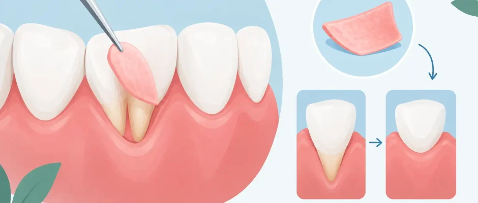
A coronally advanced flap moves the patient’s existing gum tissue toward the crown of the tooth. It can be effective when there is adequate tissue next to the recession and the flap can be positioned without tension. In thin tissue, it is frequently combined with a connective-tissue graft to improve volume and long-term stability.
A connective-tissue graft is usually harvested from the palate through a small, carefully planned access site. The graft is placed beneath the gum flap over the exposed root. It remains one of the most predictable options when the objective is both root coverage and a thicker, more resilient gum phenotype. The trade-off is a second surgical site and several days of palatal discomfort, although modern techniques and protective measures make recovery more comfortable than many patients expect.
The tunnel technique is often used when several adjacent teeth are affected, particularly in the esthetic zone. The gum is gently released beneath the surface without vertical incisions, allowing a graft or biomaterial to be inserted and the tissues to move coronally. Its advantages can include discreet scars and good blood supply, but it is technique-sensitive and not suitable for every defect.
A free gingival graft is selected primarily when the priority is to create a wider band of firm, keratinized tissue, often in the lower front region. It may not provide the same color match or level of root coverage as connective-tissue grafting, so it is chosen for function and stability rather than cosmetic perfection.
Biomaterial substitutes can reduce or eliminate the need to harvest tissue from the palate in selected cases. They may be helpful for patient comfort or when multiple sites require treatment. However, an autogenous connective-tissue graft may offer more reliable volume gain in demanding esthetic cases. The correct choice depends on biology, not on which option sounds less invasive.
What happens before and during surgery
Active gum inflammation must be controlled before periodontal plastic surgery. Professional cleaning, treatment of periodontal disease when present, and coaching on atraumatic brushing are part of the protocol. Operating in inflamed tissue compromises healing and makes the final gum margin less predictable.
The procedure is typically performed under local anesthesia. For patients with significant dental anxiety, additional sedation options can be discussed based on medical history and the surgical setting. During microsurgery, the root surface is thoroughly cleaned and conditioned when indicated, the tissue is mobilized, and the graft or substitute is stabilized with fine sutures. PRF may be used in selected cases as an adjunct to support soft-tissue healing, but it does not replace proper flap design or graft stabilization.
The appointment length varies from about one hour for a localized defect to longer for multiple teeth. Most patients leave the same day with written instructions, medication guidance when needed, and a scheduled postoperative review.
Recovery: protecting the result matters
Healing is active during the first two weeks, but tissue maturation continues for months. Mild swelling, bruising, or tenderness is possible. If tissue was taken from the palate, that area can feel similar to a superficial burn for several days. Pain is usually manageable with the postoperative regimen recommended by the surgeon.
The surgical site should not be brushed directly in the early phase. Patients typically use a prescribed antimicrobial rinse and return for professional cleaning and suture removal according to the treatment plan. Soft foods, careful chewing away from the treated area, and avoiding smoking are essential. Pulling the lip to inspect the site, brushing aggressively, or returning to vigorous exercise too early can disturb a carefully stabilized graft.
Call the clinic promptly for persistent bleeding, increasing swelling after the first few days, fever, severe pain that does not respond to prescribed medication, or a graft that appears to move. These events are uncommon, but early assessment protects the outcome.
Results, limits, and long-term stability
Predictable improvement is possible, but no ethical clinician should promise identical results for every recession defect. Complete coverage is more likely around a well-positioned tooth with preserved interdental bone, good hygiene, and adequate blood supply. Deep root grooves, extensive cervical restorations, thin tissue, active smoking, uncontrolled diabetes, and tooth movement outside the bony housing can reduce predictability.
Even after excellent surgery, the result needs maintenance. Use a soft toothbrush with gentle pressure, keep periodontal inflammation under control, attend professional hygiene visits, and wear a protective appliance if clenching or grinding is a factor. If orthodontic treatment is planned, the periodontist and orthodontist should coordinate the sequence rather than treating the gum recession in isolation.
A consultation should leave you with more than a procedure name. You should understand why the recession developed, whether it is active, what degree of coverage is realistic, where graft tissue would come from if needed, and how the result will be protected after surgery. That clarity often does more to reduce surgical anxiety than any promise of a quick fix.


