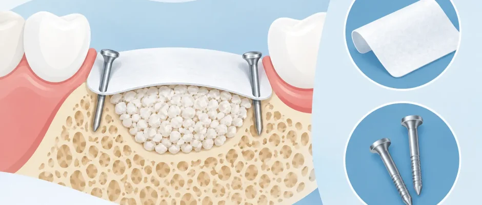
Guided Bone Regeneration: Membranes and Pins
A missing tooth does not always leave enough bone for an implant in the ideal position. The ridge may become narrow after extraction, collapse on the facial side, or lose height near the maxillary sinus or mandibular nerve. Guided bone regeneration with membranes and pins, known in Russian as «направленная костная регенерация мембрана и пины», is a surgical approach designed to rebuild that foundation before or during implant placement.
The goal is not simply to add bone material. The goal is to create and maintain a protected space where the patient’s own bone can grow predictably. For this reason, the membrane and its fixation are often as important as the graft itself.
Why bone volume matters for an implant
An implant must be surrounded by sufficient healthy bone. If the ridge is too thin, placing an implant in the available bone may force an unfavorable angle, compromise facial contour, or leave implant threads insufficiently covered. These compromises can affect hygiene, esthetics, and long-term stability.
Bone augmentation is considered when three-dimensional imaging shows a deficiency that cannot be safely or predictably managed by implant selection alone. In some cases, a narrow defect can be treated simultaneously with implant placement. In others, staged regeneration is safer: first rebuild the ridge, then place the implant after healing.
The decision depends on the defect’s size and shape, the quality of native bone, the gum phenotype, the planned tooth position, bite forces, smoking status, periodontal health, and the patient’s ability to maintain careful postoperative hygiene. A CT scan and clinical examination are essential because two sites that look similar in the mouth may require very different surgical plans.
How guided bone regeneration works
Bone has the ability to regenerate, but it needs the right conditions. Soft tissue cells from the gum grow quickly. Without a barrier, they can occupy a defect before slower-forming bone cells have time to rebuild it. A membrane separates these tissues and preserves a protected regenerative compartment.
During surgery, the area is carefully cleaned and prepared. The surgeon may use autogenous bone collected from the same surgical site, a bone substitute, or a combination. The graft acts as a scaffold and helps maintain the desired contour. A membrane is then placed over it to stabilize the material and exclude fast-growing soft tissue.
For regeneration to succeed, the graft must remain still and adequately supplied by blood from the surrounding bone and tissues. The flap must also close without tension. These principles explain why an apparently small augmentation can involve precise microsurgical technique, careful flap design, and multiple fixation points.
Resorbable and nonresorbable membranes
Resorbable collagen membranes are widely used for contained or moderate defects. They gradually break down in the body and usually do not require a separate removal procedure. Their advantages are easier handling and reduced treatment burden, but they need sufficient stability from the defect walls, graft, and surrounding tissue.
Nonresorbable membranes, including dense PTFE and reinforced options, maintain their shape longer and may be selected for larger or more demanding defects. They can provide superior space maintenance in the right indication. The trade-off is that they usually require later removal and demand especially meticulous soft-tissue closure. If exposure occurs, management becomes more complex and may reduce the regenerated volume.
There is no universally best membrane. The material should fit the anatomy, the regenerative target, the condition of the soft tissue, and the patient’s healing risk profile.
Why pins are used in bone regeneration
Membrane pins, often called tacks, are small fixation devices placed into the bone. They can secure a membrane on one or both sides of the grafted area. In selected cases, pins may also help stabilize a titanium-reinforced membrane or hold particulate graft material in the intended position.
The key benefit is immobilization. A membrane that shifts with chewing, lip movement, or flap tension may lose the protected space needed for regeneration. Even a high-quality graft material cannot compensate for instability. Pins help convert a loose covering into a stable three-dimensional framework.
Pins are particularly useful when the defect is broad, noncontained, or located on the facial aspect of the ridge, where maintaining contour is more difficult. They may also be used when a membrane must resist pressure from overlying soft tissue. In a small, well-contained defect, suturing and the natural anatomy may provide enough stability without pins.
Pin placement requires experience and planning. The surgeon must consider bone thickness, nearby tooth roots, anatomical structures, and the planned implant site. Digital imaging helps define safe fixation zones before surgery. In the posterior mandible, for example, proximity to the inferior alveolar nerve requires special attention. Technique matters more than the number of pins used.
When regeneration is performed with implant placement
Simultaneous implant placement can shorten overall treatment time when the implant achieves strong primary stability in native bone and the remaining defect can be predictably regenerated. A common example is a facial contour deficiency around an otherwise stable implant. The implant is placed according to the prosthetic plan, then graft and membrane rebuild the missing outer wall.
A staged approach is often preferable when the ridge is severely deficient, when implant stability cannot be achieved, or when the desired reconstruction requires a larger volume of bone. Staging is not a setback. It may be the more conservative way to protect the final implant position and avoid placing an implant into a compromised foundation.
In selected extraction cases, socket preservation or immediate implant placement combined with grafting can limit future collapse of the ridge. However, immediate treatment is not automatically better. Active infection, loss of socket walls, thin gum tissue, and inadequate bone for implant fixation can make a delayed protocol more predictable.
Healing and what patients can expect
After guided bone regeneration, swelling and tenderness are expected for several days. Bruising can occur, especially after larger grafting procedures. Most patients manage discomfort with a prescribed medication plan and follow-up care. The surgeon will provide instructions regarding oral hygiene, food texture, physical activity, and any temporary prosthesis.
The first two weeks are critical for soft-tissue healing. Patients should avoid brushing directly over the surgical site until instructed otherwise, avoid pulling the lip or cheek to inspect the area, and follow the prescribed rinsing protocol. Pressure from a removable denture or temporary appliance must be controlled because repeated compression can compromise the grafted area.
Bone maturation takes longer than gum healing. Depending on the procedure and materials used, implant placement after staged regeneration may be planned several months later. Follow-up examinations and imaging help confirm that healing is progressing as intended rather than relying on time alone.
Smoking and uncontrolled diabetes increase the risk of impaired healing and infection. Untreated gum disease, poor plaque control, and failure to attend follow-up visits can also threaten the result. These factors do not always rule out treatment, but they must be addressed honestly before surgery.
What makes the result more predictable
Predictable regeneration begins before the operation. Three-dimensional imaging, digital planning, and a clear restorative goal allow the surgical plan to be built around the final tooth position rather than around the bone that happens to remain. In complex cases, a surgical guide can support accurate implant positioning once the site is ready.
During surgery, atraumatic tissue handling, stable graft fixation, appropriate membrane selection, and tension-free closure work together. PRF may be used in selected cases as part of a biologically supportive protocol, particularly for soft-tissue management, but it does not replace sound grafting principles or stable fixation.
The most reassuring treatment plan is not the one that promises the fastest implant. It is the one that explains why a membrane is needed, whether pins add real value in your anatomy, and how each stage protects the final result. A careful consultation turns a complex procedure into a clear sequence of decisions, with safety and long-term function kept at the center.


