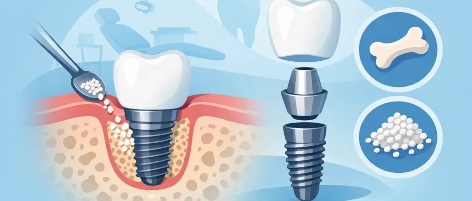
What Is Bone Graft for Dental Implants?
If you were told you need a bone graft before an implant, the question usually is not academic. It is very practical: Why can’t the implant just be placed now, and does this mean the case is becoming more serious? In simple terms, what is bone graft for dental implants? It is a procedure used to rebuild or preserve jawbone volume so an implant can be placed in a stable, safe, and prosthetically correct position.
That matters because a dental implant is not just a screw placed into any available bone. It needs enough bone in the right height, width, and quality to support long-term function and esthetics. When that foundation is missing, the surgeon may recommend bone augmentation instead of forcing a compromised implant position.
What is bone graft for dental implants and why is it done?
A bone graft is a surgical procedure that adds or stimulates new bone in an area where the jaw has become too thin or too short. The goal is not simply to “fill a hole.” The goal is to create a predictable foundation for implant placement and long-term load.
Bone loss in the jaws is common after tooth extraction, longstanding missing teeth, periodontal disease, trauma, infection, or anatomical limitations in the upper jaw near the maxillary sinus. In some patients, the bone shrinks mostly in width. In others, the issue is height. Sometimes both are reduced, and the treatment plan becomes more complex.
From a surgical standpoint, placing an implant into insufficient bone increases the risk of poor primary stability, exposure of implant threads, soft tissue problems, and esthetic compromise. In the posterior maxilla, lack of bone may also bring the sinus into play, which is why sinus augmentation is sometimes part of the plan.
When a bone graft is needed before an implant
Not every implant requires grafting. Many implants can be placed immediately after extraction or into existing healed bone without any additional augmentation. The decision depends on imaging, clinical examination, and the intended final restoration.
A bone graft may be recommended if the jawbone is too narrow for the implant diameter, if there is not enough vertical bone height, if the extraction socket has missing walls, or if a previous infection destroyed supporting bone. In the front teeth, even a mild deficiency can matter because the gum contour and smile line make small defects more visible.
This is why modern planning starts with diagnosis rather than assumptions. A CBCT scan, digital planning, and in many cases a surgical guide help determine whether grafting is necessary, how much bone is needed, and whether it can be done at the same time as implant placement or should be staged first.
How the graft actually works
Bone grafting does not mean the surgeon simply places “replacement bone” and leaves it there permanently as a block of inert material. The biology is more dynamic. The graft acts as a scaffold, and depending on the material used, it may also support the body’s own bone-forming process over time.
As healing progresses, blood supply enters the area, new bone cells populate the site, and the grafted area remodels. The exact speed and quality of this process depend on the graft material, the local blood supply, the size of the defect, surgical technique, and patient factors such as smoking, uncontrolled diabetes, or poor oral hygiene.
In guided bone regeneration, the graft is often protected with a membrane. The reason is straightforward: soft tissue heals faster than bone. Without a barrier, gum tissue can collapse into the space where bone should form. The membrane helps maintain that space long enough for bone regeneration to occur.
Types of bone grafts used in implant dentistry
Patients often ask whether the graft comes from their own body or from a manufactured source. The answer is: it depends on the clinical situation.
Autogenous bone, taken from the patient, has strong biologic potential and is still considered highly valuable in certain defects. It may be used alone or mixed with other materials. The trade-off is that it requires a donor site, which can increase surgical time and postoperative discomfort.
Xenografts and allografts are also commonly used in implant dentistry. These materials are processed and purified for medical use and are chosen because they can provide a stable scaffold and good volume maintenance in the right indication. Synthetic graft materials are another option in selected cases. None of these materials is “best” in every situation. The choice depends on the defect, the implant plan, the need for volume stability, and the surgeon’s protocol.
In many practices, biologic adjuncts such as PRF are used to support soft tissue healing and improve handling of grafted sites. This does not replace proper technique, but in the right hands it can be a helpful part of a carefully controlled surgical protocol.
Common procedures: socket graft, ridge augmentation, and sinus lift
The most conservative grafting procedure is often socket preservation after extraction. When a tooth is removed, the surrounding bone naturally begins to resorb. Placing graft material into the socket can help reduce the collapse of the ridge and make future implant placement easier and more predictable.
Ridge augmentation is performed when the jaw has already lost too much width or height. This may involve particulate graft material, membranes, tenting techniques, or block grafting in more advanced defects. These procedures are selected case by case.
In the upper back jaw, the sinus sometimes leaves too little vertical bone for implant placement. In that setting, a sinus lift may be recommended. This procedure creates space beneath the sinus membrane and places graft material to increase available bone height. In some cases the implant can be placed at the same surgery. In others, healing is allowed first.
Can the implant and bone graft be done together?
Sometimes yes. Sometimes no. This is one of the most common areas where patients hear conflicting answers, because both approaches are valid in the right scenario.
If there is enough native bone to stabilize the implant and the remaining defect can be predictably grafted around it, simultaneous implant placement may be an excellent option. It reduces overall treatment time and avoids an extra stage.
If the defect is too large, the soft tissue conditions are unfavorable, or the implant would lack primary stability, staged treatment is safer. In that case, the graft is placed first, healing is monitored, and the implant is inserted later into a more reliable foundation. Faster is not always better. Predictable is better.
Healing time and what recovery feels like
Healing after bone grafting varies with the size and type of graft. A small socket graft may heal quietly with minimal discomfort. A larger augmentation or sinus lift usually involves more swelling and a longer recovery period.
Initial healing of the gums often occurs within 1 to 2 weeks, but bone maturation takes much longer. Depending on the procedure, implant placement may be possible after a few months, or immediately if the clinical conditions are favorable. Final timing should be based on imaging and stability, not on a fixed calendar alone.
Most patients are concerned about pain. In practice, many describe grafting as easier than they expected when anesthesia, microsurgical handling, and postoperative instructions are done properly. Swelling, pressure, and temporary chewing limitations are more common than severe pain. Still, every patient heals differently.
Risks, limits, and why technique matters
Bone grafting is a routine part of advanced implant dentistry, but it is still surgery. Risks can include infection, wound opening, graft loss, membrane exposure, bleeding, sinus complications in upper jaw procedures, and incomplete regeneration. Smokers and patients with poor plaque control generally face higher complication rates.
There are also limits to what grafting can achieve. Severe defects can often be improved significantly, but biology does not always allow the same result as untouched native bone. This is why treatment planning should be realistic, especially in complex or long-neglected cases.
Technique matters as much as materials. Precise flap design, tension-free closure, stable graft fixation when needed, careful control of the soft tissue, and prosthetically driven implant planning all influence the final outcome. Digital planning and guided surgery can add another layer of accuracy, especially when bone volume is limited and implant positioning needs to be exact.
What patients should ask before agreeing to a graft
A useful consultation should leave you with clear answers to a few basic questions: Why is the graft needed in your case, what type of graft is being proposed, can the implant be placed at the same time, how long will healing take, and what risks apply specifically to your anatomy and medical history.
It is also reasonable to ask how the site will be evaluated before surgery and how success will be checked during healing. A thoughtful surgeon should be able to explain the plan in plain language without making the procedure sound casual or frightening.
For patients seeking implant treatment in complex situations, including cases with bone deficiency, this balance of clinical precision and patient comfort is exactly what makes the difference. A good plan reduces anxiety because it replaces uncertainty with a sequence you can understand.
Bone grafting is not a setback. In many cases, it is the step that makes implant treatment safer, more stable, and more esthetic in the long run. If your jaw does not currently offer enough support, rebuilding that foundation first is often the most responsible way forward.


