Your Guide to Gum Graft Surgery and Recovery
Exposed tooth roots can make a cold drink feel sharp, brushing uncomfortable, and a smile less balanced than it once was. This guide to gum graft surgery explains what the procedure can and cannot correct, how a surgeon plans it, and what a realistic recovery looks like. The goal is not simply to cover a recession defect. It is to create stable, healthy tissue around the tooth so that the root is better protected over time.
When gum recession needs surgical treatment
Gum recession means that the gum margin has moved down the root surface, exposing part of the tooth that was previously covered. It may affect one tooth or several, and it is common around canines, premolars, and lower front teeth. Recession is not always an emergency, but it should be evaluated when it is progressing, causing sensitivity, making hygiene difficult, or creating an esthetic concern.
The cause matters because surgery alone does not remove the reason recession developed. Common contributing factors include thin gum tissue, aggressive brushing, periodontal inflammation, tooth position outside the bony housing, orthodontic movement, clenching, piercings, and previous dental treatment. In some patients, a high muscle pull from the lip or cheek also places tension on the gum margin.
A graft may be recommended when the tissue is too thin to remain stable, the exposed root is sensitive or vulnerable to decay, or recession is likely to worsen. It can also be part of preparation for a restoration, orthodontic treatment, or implant-related planning. A proper assessment includes periodontal probing, evaluation of oral hygiene and bite forces, photographs, and often digital imaging. The amount of bone around the tooth and the shape of the recession determine what result is realistic.
Guide to gum graft surgery: what happens during treatment
Gum graft surgery is a microsurgical procedure performed under local anesthesia. For patients with significant anxiety, additional sedation options may be discussed depending on the clinical setting and medical history. The procedure is designed to add thickness to the existing gum and, where anatomy permits, move the gum margin toward its original position.
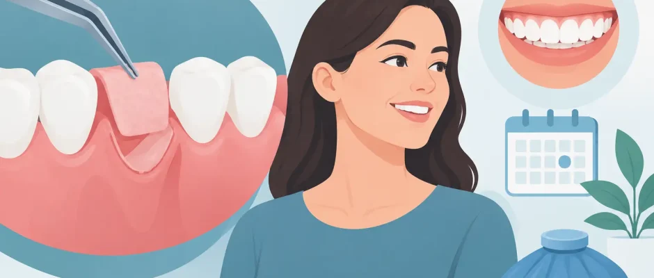
The most commonly used approaches are connective tissue grafting and tunnel-based techniques. In a connective tissue graft, the surgeon takes a thin layer of tissue from the palate or uses a suitable soft-tissue substitute in selected cases. The graft is then positioned under the gum around the recessed tooth and stabilized with fine sutures.
A tunnel technique can be especially useful when several adjacent teeth require treatment. Instead of making separate vertical incisions, the surgeon creates a precise space beneath the gum tissue, positions the graft, and advances the gum over it. This approach may reduce visible scarring and preserve blood supply, but it is not appropriate for every defect. The best technique depends on tissue thickness, recession depth, tooth position, the amount of attached gum, and the patient’s healing potential.
In certain situations, the graft is taken from the palate because the patient’s own connective tissue offers predictable biologic integration. The palate is a second surgical site, so it may cause temporary soreness. A soft-tissue substitute avoids harvesting tissue from the palate and may be useful for increasing tissue thickness, particularly over a wider area. However, it does not always provide the same degree of root coverage as an autogenous graft. This is one of the decisions where treatment must be individualized rather than standardized.
The procedure typically takes one to two hours, depending on the number of teeth and the technique. Microsurgical instruments, magnification, and delicate suturing are not cosmetic details. They help the surgeon handle tissue with less trauma and position the graft accurately, which supports predictable healing.
What results can you expect?
The desired result is healthier, thicker tissue that remains stable around the tooth. Complete root coverage is possible in many favorable cases, but it should never be promised without an examination. Deep recession, loss of bone between teeth, root prominence, cervical restorations, smoking, active inflammation, and inconsistent home care can limit the final level of coverage.
Even when full coverage is not achievable, gum grafting can still be clinically valuable. Increasing tissue thickness may reduce sensitivity, improve brushing comfort, and make the site more resistant to future recession. The result should be judged not only by the gumline position in a photograph, but by tissue stability, absence of inflammation, patient comfort, and long-term ability to maintain hygiene.
If a tooth has a non-carious cervical lesion, abrasion, or an existing filling near the gumline, the restorative plan must be coordinated with the graft. Sometimes the restoration is adjusted before surgery; in other cases, it is better to wait until the tissue has healed. Timing affects both esthetics and plaque control.
The first two weeks of recovery
Most patients describe recovery as manageable rather than severely painful. Mild swelling, tenderness, and a feeling of tightness are common during the first days. The palate may be more sensitive than the grafted area if tissue was harvested there. Prescribed medication, cold packs during the first day, and a soft diet usually help control discomfort.
The graft must remain still while new blood vessels develop. That is why the first two weeks require discipline. Do not brush or floss the surgical area until your surgeon confirms it is safe. Instead, use the prescribed antiseptic rinse exactly as directed. The rest of the mouth should still be cleaned carefully, because plaque accumulation can compromise healing.
For roughly one to two weeks, choose soft foods that do not require strong biting near the treated site. Avoid crunchy foods, seeds, hard bread crusts, spicy foods if they irritate the area, and very hot drinks during the first days. Smoking and vaping substantially impair blood supply and healing and can increase the risk of graft failure. Ideally, nicotine use should be stopped before surgery and throughout recovery.
Physical exercise can raise blood pressure and contribute to bleeding or swelling. Light activity is usually reasonable after a short rest period, while strenuous exercise should wait until the surgeon gives approval. If you wear a removable appliance or clear aligners, they must be checked before surgery so they do not put pressure on the graft.
Follow-up care protects the surgical result
Sutures are commonly removed after one to two weeks, although some may dissolve. At the follow-up visit, the surgeon evaluates graft stability, tissue color, swelling, and hygiene. It is normal for the graft to look pale or irregular initially. Its appearance changes as healing progresses, and the final tissue maturation can take several months.
Brushing is reintroduced gradually, usually with an ultra-soft brush and a specific technique. Scrubbing horizontally at the gumline is one of the most frequent reasons for recurrent recession. A clinician or hygienist may recommend a modified technique that removes plaque effectively without repeatedly traumatizing thin tissue.
Long-term maintenance includes professional periodontal monitoring, treatment of gum inflammation, control of clenching when indicated, and review of any bite-related issues. If recession developed around an orthodontically moved tooth, retainers and tooth position may also need reassessment. Surgery provides a biologic foundation, but daily habits determine whether that foundation remains stable.
Questions to ask before scheduling surgery
Before proceeding, ask what is causing the recession and whether that cause has been addressed. You should also understand which graft material is recommended, whether the procedure involves a palatal donor site, how many teeth will be treated, and what level of coverage is realistically expected in your case.
It is also reasonable to ask about the surgeon’s experience with periodontal plastic surgery, the use of magnification or microsurgical techniques, the follow-up schedule, and what is included in the treatment plan. Clear answers reduce uncertainty and help you prepare properly. In complex cases, especially when recession is combined with bone loss, implants, or previous surgery, a detailed plan is more valuable than a quick cosmetic promise.
A successful graft starts with accurate diagnosis and ends with careful maintenance. If a tooth has become sensitive, its root is increasingly exposed, or you are concerned about how the gumline is changing, an evaluation can clarify whether monitoring, hygiene changes, restorative treatment, or surgery is the right next step.


