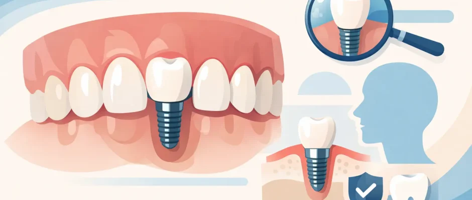
Single Tooth Implant Case Study in the Esthetic Zone
A missing front tooth changes more than a smile. It affects how a person speaks, eats, and feels in close conversation. This single tooth implant case study illustrates why replacing one tooth in the esthetic zone is not a routine “place an implant and add a crown” procedure. The goal is to restore the tooth while preserving the gum contour, bone volume, bite, and natural appearance of the adjacent teeth.
The case described below is a representative clinical scenario. Every patient requires an individual examination, CBCT scan, periodontal assessment, and restorative plan before a treatment decision is made.
Single Tooth Implant Case Study: The Clinical Challenge
A 38-year-old patient presented after trauma to an upper lateral incisor. The tooth had been treated previously, but recurrent inflammation around the root and a vertical fracture made long-term preservation impossible. The patient’s main concerns were understandable: avoiding a visible gap, keeping the gum line natural, and receiving a solution that would not compromise the neighboring healthy teeth.
An upper lateral incisor is one of the most demanding teeth to replace. The bone plate on the facial side is often thin, and even a small loss of bone or soft tissue can lead to gum recession, a dark shadow near the implant crown, or an asymmetrical smile. A technically integrated implant can still be an esthetic disappointment if the tissue architecture is not respected.
The first decision was whether immediate implant placement was appropriate. Immediate placement can reduce treatment time and help preserve tissue contours, but it is not automatically the best choice. It depends on the condition of the socket walls, the presence of infection, the available bone for primary stability, the bite, and the patient’s ability to maintain careful hygiene during healing.
Diagnosis Before Surgery
The treatment plan began with clinical photographs, digital scanning, periodontal probing, and a CBCT scan. The scan allowed three-dimensional evaluation of the root fracture, surrounding bone, nasal floor, and the position of neighboring tooth roots. It also showed that the palatal and apical bone were sufficient for implant stabilization, while the thin facial bone plate required a conservative approach.
The patient had a healthy periodontal condition overall, did not smoke, and showed no active gum inflammation around adjacent teeth. These factors supported a favorable prognosis. However, the fractured tooth had inflammation at the root apex. This meant that extraction needed to be as atraumatic as possible, with thorough cleaning of the socket before any implant could be placed.
Digital planning was used to define the ideal future crown position first. The implant was then planned in relation to that restoration, rather than simply positioned where bone appeared most available. This prosthetically driven approach helps avoid a crown that is too bulky, angled incorrectly, or difficult to clean.
Why a Surgical Guide Matters
In a single-tooth case, millimeters matter. An implant placed too far toward the lip can result in recession and an overly prominent crown. Positioned too far toward the palate, it may compromise the emergence profile or make the restoration look unnatural.
A surgical guide based on digital planning can help transfer the planned implant position to the clinical setting with greater precision. It does not replace the surgeon’s judgment. Bone quality, socket anatomy, and surgical visibility must still be assessed during the procedure. But it adds control at the stage where small deviations can affect the final result.
Atraumatic Extraction and Immediate Implant Placement
Under local anesthesia, the damaged tooth was removed using microsurgical instruments and a minimally traumatic technique. Preserving the thin facial bone and gum tissues was a priority. Excessive force during extraction can damage the socket walls and increase the need for later reconstruction.
After extraction, the socket was carefully debrided. All inflammatory tissue was removed, and the bony walls were inspected directly. The facial plate was intact, which allowed the team to proceed with immediate implant placement.
The implant was placed slightly toward the palate, following the digital plan and leaving a controlled gap between the implant surface and the facial socket wall. This positioning supports a more natural emergence profile and reduces pressure on the vulnerable facial bone.
The implant achieved adequate primary stability. Because stability was sufficient and the bite could be controlled, a temporary crown was placed on the same day. This was not a final tooth and was kept out of direct biting contact. Its purpose was to support the gum contour during healing, not to carry normal chewing forces.
A small volume of bone substitute was placed in the facial gap to support contour preservation. Depending on the defect and tissue thickness, clinicians may also use a connective tissue graft, a collagen membrane, PRF, or staged bone regeneration. In this case, the soft tissue volume was acceptable, so additional grafting was not required. That decision is always case-specific.
Healing Is Part of the Treatment, Not a Waiting Period
The first weeks after implant surgery determine much of the final soft tissue result. The patient received clear instructions: avoid biting with the temporary crown, maintain meticulous hygiene, use prescribed medications as directed, and attend follow-up visits.
Minor swelling and tenderness were expected during the first days. Significant pain, progressive swelling, fever, mobility of the temporary crown, or persistent bleeding would require prompt assessment. A calm postoperative plan helps patients distinguish normal healing from signs that need attention.
At follow-up visits, the surgical site remained stable. The gum margins around the temporary crown preserved their position, and there were no signs of infection. The patient was reminded that a temporary restoration can look close to the final result, but it should not be judged as the finished esthetic outcome. Tissue maturation takes time.
Final Restoration After Osseointegration
After approximately four months, clinical and radiographic assessment confirmed successful healing. The implant was stable, the surrounding tissues were healthy, and the bone level was maintained. A digital scan was taken to fabricate the final custom abutment and ceramic crown.
The final crown was designed to match the shape, translucency, and shade of the adjacent teeth. In the esthetic zone, color selection is not limited to choosing one shade from a guide. Natural teeth have different degrees of translucency, surface texture, and color from the neck to the incisal edge. Communication between the surgeon, restorative dentist, and dental laboratory is essential.
The crown was adjusted to avoid heavy contact during protrusive and lateral movements. This is particularly important for a lateral incisor, which can receive unfavorable sideways forces. A well-designed bite protects both the implant restoration and the natural teeth.
What Made This Result Predictable
The favorable result did not depend on one product or one surgical step. It came from a sequence of controlled decisions: accurate diagnosis, atraumatic extraction, careful cleaning of the socket, prosthetically guided implant placement, preservation of facial tissue volume, a protected temporary crown, and close follow-up.
It is equally important to recognize what could have changed the plan. If the facial bone wall had been lost, if active infection could not be fully controlled, or if the implant had not achieved primary stability, immediate placement with a temporary crown would not have been the safest option. A staged protocol with bone regeneration and delayed implant placement may take longer, but it can be the more predictable route.
What Patients Can Take From This Case
A single missing tooth can often be restored with an implant that looks and functions like a natural tooth. Yet the best result is rarely created by rushing. The treatment timeline, need for grafting, ability to receive a temporary crown, and final esthetic outcome all depend on anatomy and healing biology.
For patients considering treatment in Tel Aviv, the consultation should provide more than a price or a generic promise. It should clarify what is happening in the bone and gum tissue, whether immediate implantation is realistic, what temporary solution is appropriate, and how the final crown will be planned. At Implantolog.co.il, that discussion is built around diagnostic data and a surgical plan that respects both safety and appearance.
The right question is not simply, “Can an implant be placed today?” It is, “Which protocol gives this specific tooth the best chance of looking natural and remaining healthy years from now?”


