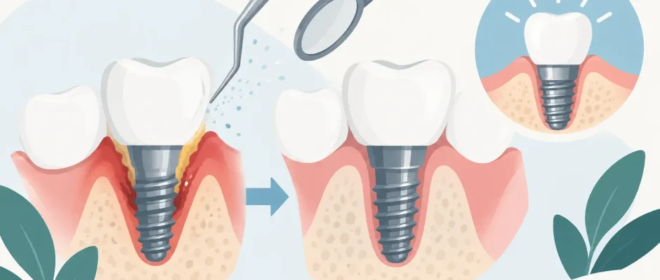
Treating Peri Implantitis to Save Your Implant
Bleeding around an implant is not a normal adjustment to be watched indefinitely. Treating peri implantitis early can stop an inflammatory process that destroys the bone supporting an implant, often before the implant becomes mobile or painful. The key is not simply cleaning the area. The treatment must identify why inflammation developed, assess how much bone has been lost, and select a protocol that gives the implant a realistic chance of long-term stability.
Peri-implantitis can progress quietly. Many patients first notice bleeding during brushing, an unpleasant taste, swelling, or a change in the appearance of the gumline. Others have no symptoms and learn about the problem during a routine examination. That is why implants require professional follow-up just as natural teeth do.
What peri-implantitis means
Peri-implantitis is inflammation of the tissues around a dental implant accompanied by progressive loss of supporting bone. It is different from peri-implant mucositis, which affects the soft tissue but has not yet caused bone loss. Mucositis is usually reversible when plaque is removed and hygiene is improved. Peri-implantitis is more serious because lost bone does not reliably return without surgical treatment, and sometimes cannot be rebuilt predictably.
The primary driver is bacterial biofilm around the implant and beneath the gumline. However, the presence of bacteria alone does not explain every case. A poorly cleansable crown contour, excess cement, residual calculus, unfavorable implant position, a history of periodontitis, smoking, uncontrolled diabetes, and irregular maintenance can all increase risk. Clenching may add mechanical overload to an already inflamed situation, although overload by itself is not considered the usual cause of peri-implantitis.
A reliable plan begins with diagnosis rather than assumptions. The examination includes gentle probing around the implant, assessment of bleeding or suppuration, mobility testing, evaluation of the bite and prosthetic design, and comparison of current radiographs with previous images. Three-dimensional imaging may be useful when the pattern and extent of bone loss are unclear or when regenerative surgery is being considered.
Treating peri implantitis starts with the cause
There is no universal procedure that is correct for every implant. An implant with shallow bone loss and accessible surfaces may respond to nonsurgical decontamination and a strict maintenance program. A deeper crater-like defect may require surgery to gain visibility, clean the implant surface thoroughly, and possibly regenerate bone. If the implant is severely mobile or the remaining bone support is inadequate, removal may be the safer and more predictable choice.
This conversation can feel discouraging, but it is an essential part of responsible care. The goal is not to promise that every implant can be saved. The goal is to preserve a treatable implant when prognosis is favorable and avoid repeated procedures when removal and a carefully planned replacement would provide a better outcome.
Step one: control inflammation and improve access
Initial treatment usually focuses on reducing bacterial load and making daily cleaning possible. Professional instruments designed for implants are used to remove deposits while minimizing damage to the implant surface. Air-polishing with low-abrasive powder, ultrasonic devices with appropriate tips, and antiseptic protocols may be part of the approach, depending on the clinical situation.
Equally important is the restoration above the implant. A crown or bridge that leaves no room for an interdental brush, traps food under its contour, or sits too deeply below the gumline may prevent successful hygiene. In some cases, modifying or temporarily removing the restoration is necessary to properly clean and inspect the implant.
Antibiotics are not a substitute for biofilm removal. They may be considered in selected cases, particularly with acute infection or as an adjunct to surgical treatment, but they are not routinely prescribed simply because bleeding is present. Unnecessary antibiotics can mask a problem without resolving it and contribute to resistance.
Step two: reassess the tissue response
After initial therapy, the tissues need time to heal before a final decision is made. Reduced bleeding, shallower probing depths, improved home care, and stable radiographic findings are encouraging signs. Persistent bleeding, pus, deep pockets, or progressive bone loss indicate that nonsurgical care alone is unlikely to be sufficient.
This reassessment is where individualized planning matters. The location of the implant, esthetic demands, bone defect shape, health conditions, smoking status, and ability to attend maintenance visits all influence the recommended next step.
When surgical treatment is needed
Surgery provides direct access to the contaminated implant surface and the bone defect. Under local anesthesia, the gum tissue is carefully lifted, inflammatory tissue is removed, and the implant threads are decontaminated using a protocol selected for the implant surface and defect anatomy. The restoration may need to be removed or adjusted to make this treatment effective.
There are two main surgical strategies. Resective treatment reshapes the bone and soft tissue to create a contour that is easier to clean and maintain. It can be effective in non-esthetic areas or when bone regeneration is not predictable. The trade-off is that the implant surface may become more exposed, so meticulous hygiene becomes even more important.
Regenerative treatment aims to rebuild a contained bone defect using grafting material, often combined with a barrier membrane. PRF, prepared from the patient’s own blood, may be used as a biologic adjunct in selected cases to support soft-tissue healing. Regeneration is most promising when the defect has favorable walls that can contain the graft, the implant is stable, and infection control is achievable. It is not a cosmetic procedure and cannot guarantee complete restoration of the original bone level.
Modern planning may include digital scans, radiographs, and three-dimensional imaging to assess the defect before surgery. These tools improve precision, but they do not replace clinical judgment. The best technology is useful only when it supports a clear surgical objective and a realistic prognosis.
When removing the implant is the better decision
An implant that moves is generally considered to have lost osseointegration and usually cannot be saved. Removal may also be recommended when bone loss is extensive, inflammation repeatedly returns despite appropriate treatment, implant position prevents hygienic restoration, or the surrounding bone anatomy makes predictable regeneration unrealistic.
Removal is not the end of treatment. The area can be allowed to heal, with or without bone preservation depending on the defect. A new implant may be planned later if there is sufficient regenerated bone, healthy soft tissue, and a design that allows proper cleaning. In more complex cases, staged bone augmentation or a different prosthetic solution may be safer than immediate reimplantation.
For patients who have already experienced one implant complication, a slower, carefully staged plan often provides more value than rushing toward another implant placement.
What protects the result after treatment
Successful treatment continues at home and during maintenance appointments. Brushing alone may not clean the spaces around implants, especially under bridges. The most appropriate tools may include an interdental brush of the correct size, implant-specific floss or tape, and an oral irrigator as an adjunct. The exact routine depends on the restoration design and should be demonstrated in the mouth, not explained only in general terms.
Professional maintenance intervals are individualized. A patient with previous periodontitis, a history of peri-implantitis, smoking exposure, or difficult-to-clean restorations often benefits from shorter recall intervals. At these visits, the clinician monitors bleeding, probing depths, plaque control, bite forces, and radiographic bone levels when indicated.
Smoking cessation and diabetic control are not side issues in this process. They directly affect healing, inflammation, and the predictability of surgical treatment. If clenching is present, a night guard and bite adjustment may reduce mechanical stress on the restoration and implant complex.
Do not wait for pain
Peri-implantitis is often manageable, particularly when identified before severe bone loss occurs. But it is not a condition to treat with repeated mouthwash use or by waiting to see whether bleeding disappears. A focused clinical examination can clarify whether the issue is superficial mucositis, an early bone defect, a restoration-related problem, or a more advanced infection requiring surgery.
If you notice bleeding, swelling, discharge, bad taste, or loosening around an implant, arrange an evaluation promptly. A calm, structured plan is usually far more effective than emergency treatment after the implant has lost critical support.


