Best Methods of Bone Grafting in Implantology
When a patient is told, “You do not have enough bone for an implant,” the real question is not whether bone grafting exists. The real question is which approach gives the safest and most predictable result in this specific anatomy. That is exactly why the topic of the best methods of bone grafting in implantology cannot be reduced to a ranking. In implant surgery, the best method is the one that matches the defect, the soft tissue condition, the implant plan, and the patient’s healing profile.
What makes the best methods of bone grafting in implantology
Bone augmentation is not one procedure. It is a group of surgical techniques used to rebuild lost volume so an implant can be placed in a prosthetically correct and stable position. The main variables are simple: how much bone is missing, in which direction it is missing, whether the site is infected or healed, and whether implant placement should happen immediately or in stages.
A narrow ridge, a vertical defect, and a posterior upper jaw with sinus expansion are three very different problems. They should not be treated as if one method fits all. Good planning starts with CBCT imaging, an evaluation of soft tissue thickness, occlusion, and the final restoration design. If the prosthetic plan is weak, even technically successful grafting can produce a compromised result.
Best methods of bone grafting in implantology by clinical scenario
Guided bone regeneration for small to moderate defects
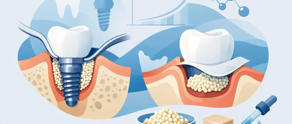
Guided bone regeneration, or GBR, is often the workhorse of modern implantology. It is commonly used when the ridge is too thin or when the implant will have exposed threads after placement. The principle is straightforward: the surgeon places graft material in the deficient area and protects it with a membrane so bone can regenerate while soft tissue is kept out of the space.
For many horizontal defects, GBR offers an excellent balance between effectiveness and morbidity. It is less invasive than harvesting a large block graft and is highly versatile. It can be done simultaneously with implant placement in selected cases, which shortens treatment time.
The trade-off is that GBR is technique-sensitive. Stability of the graft, membrane choice, tension-free closure, and infection control all matter. If the soft tissue is thin or closure is under tension, the risk of wound opening increases, and with it the risk of partial graft loss. In experienced hands, GBR is one of the strongest options for predictable contour augmentation.
Sinus lift for the posterior upper jaw
When bone is missing in the upper back jaw, the issue is often not just bone loss after extraction. The maxillary sinus gradually expands, leaving limited vertical height for implants. In this situation, sinus augmentation is frequently the right answer.
There are two main approaches. A crestal sinus lift is usually considered when only a modest gain is needed. A lateral window sinus lift is used when the residual bone is very limited or when more substantial augmentation is required. Neither is automatically “better.” The decision depends on residual height, sinus anatomy, membrane condition, and implant stability.
Sinus lifting has a long track record and very good outcomes when indications are respected. The main nuance is that sinus surgery is anatomy-dependent. Septa, membrane thickness, previous inflammation, or sinus pathology can change the plan. This is where preoperative imaging and a surgeon comfortable with complex anatomy become especially important.
Autogenous block grafts for severe ridge deficiency
For significant horizontal and especially vertical loss, autogenous bone blocks still have an important role. These grafts are taken from the patient’s own bone and fixed to the deficient site to rebuild volume that particulate grafting alone may not reliably create.
Why are they still used despite the rise of biomaterials? Because in larger defects, especially when vertical gain is needed, the patient’s own cortical-cancellous bone can offer strong biological potential and structural support. In other words, there are cases where less invasive methods may look attractive but create a compromise in implant position or esthetics.
The downside is obvious: this is a more invasive procedure. It may require a donor site, more postoperative swelling, and a longer healing phase. It also demands precise fixation and contour management. For the right case, though, it can turn a poor foundation into a restorable site instead of forcing a prosthetic workaround.
Ridge preservation after extraction
Some of the best bone grafting is done before a major defect develops. Ridge preservation is performed at the time of tooth extraction to reduce the collapse of the socket walls and maintain future implant options. It does not freeze the ridge in time, but it can significantly limit bone loss.
This matters because a well-managed extraction socket can reduce the need for larger augmentation later. Not every extraction site requires the same protocol. The amount of infection, the condition of the buccal plate, and whether immediate implant placement is planned all affect the decision.
For patients, this is one of the most practical points to understand: timing changes everything. A tooth removed without site preservation may later require a more complex reconstruction than a tooth removed with a careful regenerative plan.
PRF as a biologic adjunct, not a replacement
PRF, or platelet-rich fibrin, is often discussed as if it were a stand-alone solution. It is not. PRF is a biologic adjunct that may support healing, soft tissue quality, and graft maturation. It can be useful in extraction sites, GBR, sinus procedures, and soft tissue management.
Its value is real, but it should be described honestly. PRF does not replace bone where substantial volume is missing, and it does not eliminate the need for sound surgical technique. Used appropriately, it improves the biologic environment. Used as marketing language without indication, it creates unrealistic expectations.
How surgeons choose between graft materials
Patients often ask which graft material is best: your own bone, donor bone, bovine-derived mineral, or synthetic substitutes. The honest answer is that each has strengths and limitations. Autogenous bone has strong biologic activity but resorbs faster and requires harvesting. Xenograft materials are often excellent for maintaining volume over time but remodel more slowly. Allografts and synthetics can be effective in the right indication and are often part of combination protocols.
The key is not brand preference. It is defect biology. In some cases, mixing materials makes sense because one component supports regeneration while another helps preserve contour. In esthetic areas, long-term volume stability may matter more than speed. In other cases, faster turnover is desirable.
When immediate implant placement works – and when it does not
Patients understandably like the idea of extraction, grafting, and implant placement in one appointment. Sometimes this is ideal. Sometimes it is the wrong move.
Immediate implant placement can preserve treatment time and reduce the number of surgeries, but it works best when infection is controlled, primary stability is achievable, and the implant can be placed in a prosthetically correct position with adequate surrounding bone. If the facial plate is missing or the soft tissue is compromised, staging the procedure may produce a safer and more esthetic result.
This is one area where experience matters more than speed. Fast treatment is valuable only when it does not sacrifice tissue stability.
What patients should look for beyond the method itself
The discussion about the best methods of bone grafting in implantology should always include execution. A good method can fail with poor planning, and a demanding case can succeed with disciplined protocol. Three things matter more than patients are often told.
First is diagnosis. CBCT-based planning is not a luxury in complex implant cases. It is the basis for understanding the true defect and avoiding guesswork. Second is soft tissue management. Bone grafting is often judged by the graft, but closure, flap design, and tension control frequently determine whether the result holds. Third is prosthetic planning. Bone should be rebuilt for the final tooth position, not simply to fill a void.
In advanced practices, digital planning and surgical guides can improve precision, especially when augmentation and implant positioning must work together. That does not replace surgical judgment, but it does improve predictability.
The most honest answer: the best method depends
Patients usually want a single answer. Surgeons owe them a precise one. Small horizontal deficiency around an implant site may be best treated with GBR. Severe vertical loss may call for a block graft or staged reconstruction. Posterior maxillary atrophy often points to sinus augmentation. A fresh extraction socket may benefit most from ridge preservation performed well and on time.
The best treatment plan is not the one with the most technology or the shortest timeline. It is the one that creates a stable foundation for the implant, respects anatomy, reduces risk, and supports long-term function and esthetics. In a field where bone volume, soft tissue, and implant position are tightly connected, careful planning is part of the surgery.
If you have been told that you lack bone for implants, that is not the end of the conversation. It is the point where the right surgical strategy starts to matter.
Comments (0)