A Complex Bone Graft Case Study in Implant Care
A patient may hear, “There is not enough bone for an implant,” and assume that the answer is simply no. In a complex bone graft case study, the more useful question is different: why is bone missing, where is it missing, and what treatment sequence can rebuild support without creating unnecessary risk?
This is the type of decision that cannot be made from a panoramic X-ray alone. Implant treatment in an area of advanced bone loss requires three-dimensional diagnosis, an assessment of the gums and bite, and a plan that respects both biology and the patient’s priorities. The goal is not merely to place an implant. It is to create stable conditions for a restoration that can function comfortably for years.
The clinical scenario below is representative and anonymized. Every patient requires an individual examination and treatment plan.
The Clinical Challenge: Long-Standing Tooth Loss
A 54-year-old patient presented with a missing upper first molar. The tooth had been extracted several years earlier after repeated inflammation and unsuccessful restorative treatment. The patient wanted a fixed replacement and was concerned that a removable solution would feel uncomfortable.
At first glance, the space appeared suitable for an implant. However, the cone beam CT scan showed two significant limitations. The height of native bone beneath the maxillary sinus was limited, and the ridge had become narrow after years without a tooth. The sinus membrane was healthy, but the available bone did not provide a predictable foundation for a standard implant placed immediately.
This distinction matters. An implant may sometimes be inserted into limited bone, but achieving initial stability is not the same as obtaining a durable, biologically sound result. Placing an implant too short, too narrow, or at an unfavorable angle simply to avoid grafting can compromise the future crown, hygiene access, and load distribution.
The patient also had a history of nighttime clenching. That did not rule out treatment, but it increased the importance of correct implant position, adequate bone support, and a protective night guard after restoration.
Complex Bone Graft Case Study: Diagnosis Before Surgery
The planning process began with a clinical examination, periodontal assessment, digital scan, and cone beam CT. These records allowed the future crown position to guide the surgery rather than allowing the remaining bone alone to determine where the implant would be placed.
The key findings were clear: approximately 4 to 5 mm of bone height was present below the sinus in the intended implant area, while the ridge width was insufficient for an ideally positioned implant without augmentation. The neighboring teeth were stable, and there was no active gum infection. This made a regenerative approach reasonable.
Several paths were discussed with the patient. One option was a short implant, if the ridge dimensions and bite allowed it. Another was a bridge supported by the adjacent teeth. A third option was staged bone reconstruction followed by implant placement and a single crown.
There is no universal “best” solution. A short implant can be an excellent treatment in selected cases, particularly when adequate ridge width and favorable occlusion are present. A bridge may be appropriate when surgery is not desired or when neighboring teeth already require crowns. In this case, the patient preferred to preserve the adjacent healthy teeth and choose a treatment plan designed around a prosthetically ideal implant position.
Why a Staged Approach Was Chosen
A simultaneous sinus lift and implant placement can reduce treatment time when enough native bone is available for reliable primary stability. In this case, the combination of reduced bone height and ridge deficiency made that approach less predictable.
A staged protocol was selected for safety and control. The first surgery would rebuild bone volume and elevate the sinus floor. Implant placement would follow only after the regenerated site demonstrated appropriate healing.
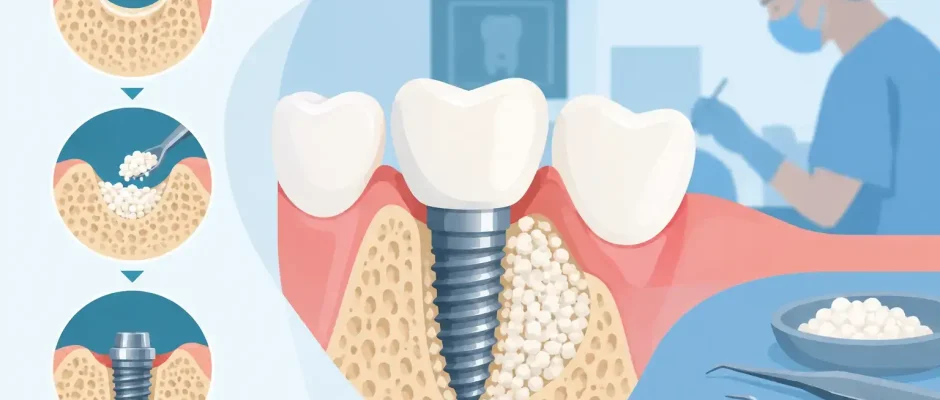
The procedure included lateral sinus floor elevation with careful management of the sinus membrane. A particulate graft material was placed to maintain space for new bone formation. Because the ridge also needed horizontal support, guided bone regeneration was performed using graft material and a barrier membrane to protect the regenerative area from soft-tissue ingrowth.
The graft is not a substitute for the patient’s own healing. Its role is to maintain volume and provide a scaffold while the body gradually forms and remodels bone. The membrane is equally important in selected defects because it helps protect that space during early healing.
PRF was used as an adjunct to support soft-tissue healing and handling of the surgical site. PRF is not a magic ingredient and does not replace correct graft stabilization or atraumatic technique. Used appropriately, it can be a valuable part of a biologically focused protocol.
The Surgical Day: Precision and Patient Comfort
For the patient, a complex grafting procedure should not feel mysterious. Before surgery, the sequence, expected swelling, pain-control plan, dietary restrictions, and follow-up schedule were reviewed in plain language.
Local anesthesia was used to ensure comfort during the procedure. The surgery was performed with careful flap design and microsurgical tissue handling, because the quality of soft-tissue closure affects graft protection. The graft and membrane were stabilized, the sinus membrane was checked, and tension-free closure was achieved with sutures.
Patients often worry most about pain. In reality, swelling, nasal precautions after sinus surgery, and temporary dietary adjustments are often more relevant during the first postoperative days. Discomfort is usually managed with the prescribed medication plan, cold compresses during the initial period, and clear instructions on what is normal and what requires a call to the clinic.
For a sinus graft, patients are generally asked to avoid forceful nose blowing, closed-mouth sneezing, smoking, and strenuous activity during the early healing phase. These instructions are not formalities. Pressure changes and smoking can interfere with healing and increase complication risk.
Healing Is an Active Part of Treatment
The patient returned for postoperative examinations to evaluate soft-tissue healing and remove sutures. There were no signs of infection, graft exposure, or sinus complications. The patient followed the recommendations carefully, including temporary modifications to diet and physical activity.
Bone regeneration cannot be judged by how the site looks from the outside. After an appropriate healing period, a follow-up CBCT scan was obtained. It showed increased vertical bone volume beneath the sinus and sufficient horizontal ridge width for a properly sized implant in the planned restorative position.
This is one reason staged care can be reassuring in demanding cases. It creates a checkpoint. If healing is incomplete or the anatomy changes, the plan can be adjusted before an implant is placed. Moving forward is based on clinical evidence, not optimism.
Implant Placement and Prosthetic Planning
Once adequate bone was confirmed, the implant was placed using a digitally planned position. A surgical guide can be particularly helpful in reconstructed areas because the available volume may be limited and the desired crown position is precise. The guide does not replace surgical judgment, but it supports accurate translation of the digital plan to the operating field.
Primary stability at placement was satisfactory. The implant was allowed to integrate before the final crown was made. During this phase, the restorative plan considered the patient’s bite and clenching history. The final crown needed contact points that were easy to clean, a contour that supported the gum tissue without pressure, and occlusion designed to reduce excessive lateral loading.
After restoration, the patient received instructions for daily cleaning around the implant and was advised to use a night guard because of clenching. An implant is not protected from gum inflammation simply because it cannot decay. Plaque control, professional maintenance, and bite management remain essential.
What This Case Shows – and What It Does Not
The favorable result in this complex bone graft case study came from sequencing, not from one material or one surgical maneuver. Three-dimensional diagnosis identified the real limitations. Regeneration addressed the missing foundation. Implant placement occurred only after the site was ready, and the crown was planned as part of the surgical decision from the beginning.
At Implantolog.co.il, complex implant cases are approached with that same discipline: diagnostics first, a clear explanation of options, and treatment selected for long-term predictability rather than speed alone.
Bone grafting is not necessary for every patient with missing teeth. It may be avoidable with a short implant, an angled implant, a different implant position, or a non-implant restoration. Conversely, when reconstruction is indicated, skipping it can be the more costly compromise.
The most helpful next step is a consultation with clinical examination and 3D imaging. A clear plan can turn “not enough bone” from a discouraging statement into a manageable path toward comfortable function and a stable smile.


