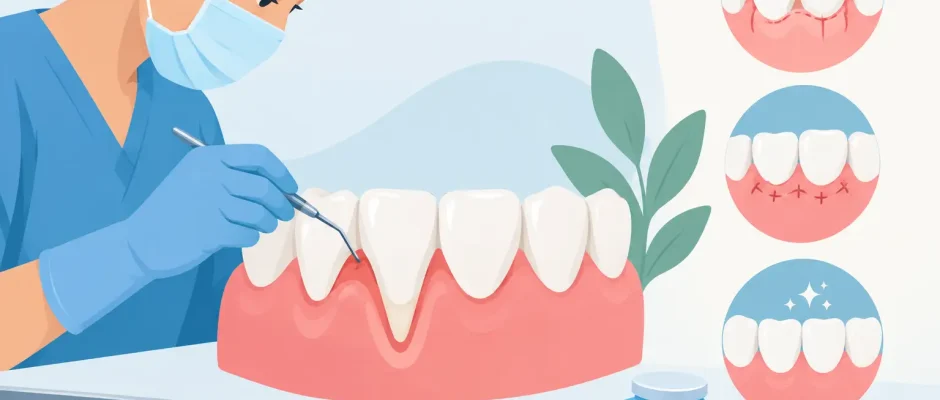
Exposed root surfaces can make a tooth feel suddenly vulnerable: cold water causes a sharp sensation, the gumline looks uneven, and brushing becomes uncomfortable. This guide to gum recession surgery explains when coverage surgery is appropriate, what the procedure involves, and how a carefully planned microsurgical approach can protect the tooth for the long term.
Gum recession is not only an esthetic concern. As the gum margin moves away from the crown, root cementum becomes exposed. This surface is softer than enamel, more prone to wear and decay, and can be sensitive to temperature or touch. Recession may affect one tooth or several teeth, often around lower front teeth, canines, or teeth that sit slightly outside the dental arch.
Why gums recede and why the cause matters
A graft can improve tissue thickness and root coverage, but surgery alone does not remove the reason recession developed. The first task is to identify the contributing factors. These may include thin gum tissue, naturally prominent roots, aggressive horizontal brushing, periodontal inflammation, orthodontic tooth movement, clenching, or a high frenum pull on the lip.
In some cases, recession occurs around a tooth with otherwise healthy bone and stable periodontal support. In others, it is part of active periodontal disease, where inflammation has already caused loss of bone and attachment. These situations require different treatment sequences. Active inflammation must be controlled before elective root coverage surgery is considered.
A clinical examination evaluates plaque control, bleeding, pocket depth, tooth mobility, bite forces, the width and thickness of keratinized tissue, and the position of the tooth within the bone. Periapical X-rays, and in selected complex cases 3D imaging, help assess bone support and rule out root fractures, infection, or other pathology. A predictable plan begins with a diagnosis, not with choosing a graft technique by name.
Guide to gum recession surgery: when it is recommended
Surgery is commonly considered when exposed roots are causing sensitivity, progressive recession, root wear, esthetic asymmetry, or difficulty maintaining hygiene. It may also be advised before certain restorative or orthodontic procedures when the existing tissue is too thin to remain stable.
Not every recession defect needs surgery. If the recession is stable, there is no sensitivity, and cleaning is straightforward, regular monitoring and a gentler hygiene protocol may be the most reasonable approach. Conversely, waiting can be a poor trade-off when the defect is deepening, the root is wearing away, or the patient has very thin tissue at a vulnerable tooth.
The likelihood of complete root coverage depends on several clinical details: the amount of interdental bone and papilla support, the depth and width of the recession, tissue thickness, tooth position, smoking status, and the patient’s ability to follow aftercare instructions. A responsible consultation should include an honest discussion of what can be improved, rather than a promise that every root can be covered completely.
How root coverage surgery is performed
Most gum recession procedures are completed under local anesthesia. For patients with significant dental anxiety, additional comfort measures can be discussed in advance. The goal is not simply to move gum tissue upward. The surgeon creates a stable, well-vascularized environment in which the tissue can heal in a higher, thicker position.
Connective tissue graft
A connective tissue graft remains one of the most predictable options, especially for thin gum biotypes and isolated recession around visible teeth. A small layer of connective tissue is usually obtained from the palate and placed beneath a carefully elevated gum flap at the treatment site. The flap is then positioned to cover the graft and secured with fine sutures.
This technique provides both root coverage and an increase in tissue thickness. The trade-off is that there are two healing areas: the recipient site and the palate. Modern harvesting techniques and protective dressings can make palatal recovery more comfortable, but patients should expect temporary tenderness.
Tunnel technique
The tunnel technique is a microsurgical approach often used for one or multiple adjacent recession defects. Rather than making broad vertical incisions, the surgeon prepares a space beneath the gums and inserts a connective tissue graft through a small access point. The gum margin is then advanced and stabilized with delicate sutures.
Because the papillae and blood supply are preserved as much as possible, this method can offer very natural esthetic healing. It is technically demanding and is not suitable for every anatomy, but it is particularly valuable in the smile zone when tissue handling must be precise.
Coronally advanced flap and alternatives
When there is sufficient gum thickness and favorable anatomy, a coronally advanced flap may be used with or without a graft. The existing gum tissue is gently released and repositioned over the exposed root. In selected cases, biologic materials or a collagen matrix can be considered instead of tissue from the palate.
These alternatives can reduce discomfort at the donor site. However, they may not provide the same increase in tissue thickness or the same long-term stability in every high-risk defect. The correct choice depends on the site, the esthetic goal, and the need for durable tissue volume.
What happens before surgery
The surgery itself is only one stage of treatment. Before the procedure, professional cleaning may be needed to reduce inflammation. Brushing technique is reviewed carefully, usually with a soft brush and minimal pressure at the gumline. If a sharp tooth edge, poorly contoured restoration, or bite interference is traumatizing the tissue, it should be addressed as part of the plan.
Photographs and measurements document the starting position of the gumline. This allows the patient and surgeon to evaluate healing objectively rather than relying only on memory. For complex esthetic cases, the treatment plan may also involve the restorative dentist or orthodontist.
Smoking and nicotine use increase the risk of impaired blood supply and less predictable healing. Patients should discuss this openly. It may be appropriate to postpone surgery until nicotine exposure is stopped, particularly when extensive grafting is planned.
Recovery after gum graft surgery
The first week is about protecting the surgical site. Mild swelling, bruising, and tightness are possible, while significant worsening pain, fever, pus, or uncontrolled bleeding require prompt contact with the treating clinic. Most patients can return to routine nonphysical work quickly, but strenuous exercise is generally avoided for several days because it can increase bleeding and swelling.
For the initial healing period, patients should not brush or floss directly over the grafted area. A prescribed antiseptic rinse may be used instead. Food should be soft, cool to lukewarm, and easy to chew away from the surgical site. Yogurt, eggs, soft fish, pasta, and cooked vegetables are often more comfortable than crusty bread, seeds, nuts, or spicy foods.
Fine sutures are usually checked or removed according to the clinical protocol, often within one to two weeks. The tissue may look fuller, lighter in color, or slightly uneven during early healing. This is normal. The final contour and color blend develop gradually over several months.
A graft is vulnerable to repeated trauma in the early phase. Pulling the lip to inspect the site, brushing too soon, chewing hard food on the area, or smoking can compromise healing. Following instructions precisely is not a minor detail. It directly affects the stability of the surgical result.
Risks, limits, and realistic expectations
Gum recession surgery has a strong record when it is correctly indicated and performed with careful tissue management, but it is still surgery. Possible complications include bleeding, infection, swelling, prolonged sensitivity, graft shrinkage, incomplete root coverage, and recurrence of recession over time.
Complete coverage is more likely when the papillae and bone between teeth are intact. If there has been substantial loss of interdental support, the objective may be to thicken and stabilize the tissue, reduce sensitivity, and improve appearance rather than achieve a perfectly even gumline. That can still be a meaningful clinical result.
Long-term success also depends on maintenance. A graft does not make a tooth immune to inflammation, traumatic brushing, or periodontal disease. Gentle daily hygiene, periodontal checkups, and management of grinding or bite-related trauma are part of protecting the investment in treatment.
Choosing the right surgical plan
The best technique is not the newest procedure or the one with the shortest recovery. It is the method that matches the anatomy of the defect and the patient’s goals. A microsurgical approach, magnification, fine sutures, and careful flap design can improve precision and patient comfort, but they must be paired with sound diagnosis and disciplined aftercare.
For patients considering treatment in Tel Aviv, a consultation with a surgeon experienced in periodontal esthetic surgery should result in a clear plan: which teeth require treatment, whether a graft is necessary, what level of coverage is realistic, and how healing will be monitored. Clarity before surgery is often the first step toward feeling calm about it.
A healthy, stable gumline is built gradually. When sensitivity, visible root exposure, or continuing recession is addressed early and with a tailored protocol, surgery can protect more than the appearance of a smile – it can help preserve the tooth itself.


