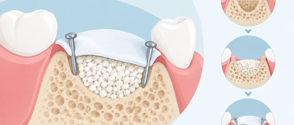
When an implant site lacks bone on the facial side, placing an implant immediately can mean accepting an unstable foundation or an esthetic compromise. This clinical example of bone regeneration with a membrane and pins shows why guided bone regeneration is often planned as a separate, highly controlled stage before implant placement.
The goal is not simply to make an X-ray look better. The surgeon must create and maintain enough three-dimensional bone volume to support the implant, preserve the soft-tissue contour, and allow predictable oral hygiene over many years. A membrane and fixation pins are used when the graft needs reliable protection from pressure and movement during healing.
The clinical problem: a narrow ridge after tooth loss
Consider a patient who lost an upper lateral incisor after a fracture and infection. The tooth had been removed elsewhere, and several months later the area had healed without active inflammation. On examination, the gum appeared healthy, but the ridge was visibly and palpably narrow.
A CBCT scan confirmed the concern: there was enough vertical height for an implant, but insufficient facial bone width. In the anterior maxilla, this distinction matters. An implant may technically fit into the available bone, yet be positioned too far toward the palate or have an excessively thin facial bony wall. Both situations can affect the final gum contour and long-term esthetics.
The treatment plan was therefore staged. First, guided bone regeneration would rebuild the missing facial contour. After maturation of the graft, a second CBCT scan would confirm whether implant placement could be performed in the ideal prosthetic position.
Example of bone regeneration with a membrane and pins
After local anesthesia, the surgical area is accessed through a carefully designed flap. In an esthetic zone, flap design is not a minor detail. It must provide visibility and tension-free closure while respecting the blood supply and preserving as much soft tissue as possible.
The recipient bone is then cleaned of fibrous tissue and evaluated directly. Small cortical perforations may be created when indicated to encourage bleeding from the marrow spaces and access to regenerative cells. The graft material is placed where the ridge volume is deficient, with deliberate overbuilding only when the anatomy and soft-tissue envelope permit it.
A resorbable collagen membrane is shaped to cover the graft completely. The membrane acts as a barrier: it helps prevent faster-growing soft-tissue cells from occupying the space where bone formation is intended to occur. But a membrane is effective only when it remains stable. If it collapses into the graft or shifts during healing, the protected space is lost.
This is where fixation pins become important. Small titanium pins can secure the membrane to the native bone on the palatal and facial sides. They prevent micromovement, help maintain the desired contour, and reduce the risk that flap pressure displaces the graft. In larger horizontal defects, stable fixation is often more predictable than relying on sutures alone.
The flap is then advanced and closed without tension using fine sutures. Tension-free closure is essential. A graft can be well positioned and a membrane perfectly fixed, yet still be compromised if the wound margins are under excessive strain and open during the early healing period.
Why pins are not used in every case
Membrane fixation is a tool, not a routine requirement for every graft. A very small contained defect, especially one surrounded by intact bony walls, may be treated successfully with a different stabilization approach. Conversely, a broad facial defect, a vertical deficiency, or a non-contained defect usually demands more rigid control of the grafted space.
The decision depends on CBCT findings, defect morphology, soft-tissue thickness, implant timing, and the intended final restoration. The surgeon also considers whether a resorbable membrane is appropriate or whether a non-resorbable reinforced membrane would provide better structural support. Each option has trade-offs in handling, healing, and the need for a possible second procedure.
Healing: what the patient can expect
For the first several days, swelling and mild bruising are possible. Discomfort is typically controlled with the prescribed medication plan, cold compresses during the first day, and a soft diet. The patient should avoid brushing directly over the operated area until instructed otherwise and should not pull the lip to inspect the wound repeatedly.
Mechanical protection matters as much as medication. Pressure from a removable temporary appliance, trauma from brushing, smoking, or early heavy chewing can disturb the surgical site. Smoking in particular reduces blood supply and increases the risk of wound healing complications. For a procedure designed to regenerate tissue, this is a meaningful risk rather than a minor lifestyle recommendation.
Follow-up visits allow the surgeon to assess tissue healing, remove sutures when appropriate, and identify early signs of membrane exposure or infection. Most bone grafts require several months of maturation before implant placement. The exact interval depends on the original defect, graft material, the patient’s healing capacity, and the stability achieved during surgery.
What makes this approach predictable
Guided bone regeneration succeeds through details that are easy to underestimate. A stable graft, adequate blood supply, exclusion of unwanted soft tissue, protected space, and primary wound closure work together. If one of these elements is compromised, the result may be less volume than planned.
Digital planning improves decision-making before surgery. CBCT imaging shows the shape of the defect in three dimensions, while intraoral scans and prosthetic planning help determine where the future tooth should emerge. This is especially valuable in the smile zone: the graft should be planned around the final tooth position, not merely around where bone appears easiest to add.
In selected cases, platelet-rich fibrin, or PRF, may be incorporated into the protocol to support soft-tissue healing. PRF does not replace membrane fixation or sound surgical technique, but it can be a useful biologic adjunct when chosen for the right indication.
Risks and realistic expectations
Even with careful planning, bone regeneration is surgery and carries risks. These include postoperative swelling, bleeding, infection, wound opening, partial graft resorption, membrane exposure, and the possibility that additional grafting may be needed before implant placement. Pins may also need to be removed if they remain exposed or interfere with a later procedure.
A successful graft does not mean that every patient receives the same amount of new bone or follows the same timeline. Healing is influenced by smoking, diabetes control, periodontal health, oral hygiene, medications, previous infection, and the condition of the surrounding soft tissue. In some cases, a staged approach is safer than attempting extraction, grafting, and implant placement on the same day.
The most responsible plan is one that explains these variables before treatment begins. A patient should understand not only what will be done, but why a particular technique has been selected and what alternatives are reasonable.
From regenerated bone to implant placement
After the healing phase, a new clinical examination and, when indicated, CBCT scan assess the reconstructed ridge. The surgeon evaluates width, contour, and density, then plans implant placement according to the final crown position. If the regenerated volume is sufficient, the implant can be placed with appropriate facial bone support and a more favorable emergence profile.
For patients, the value of this staged process is often not obvious at the first consultation. It can feel slower than placing an implant immediately. Yet when the foundation is deficient, taking time to reconstruct it may reduce compromises in implant position, gum architecture, and long-term esthetics.
A membrane and pins are small components of a larger surgical protocol, but they can make a decisive difference when a graft must hold its shape. The right next step is a clinical examination and three-dimensional imaging that show whether your case needs regeneration, immediate implantation, or a different path altogether.


