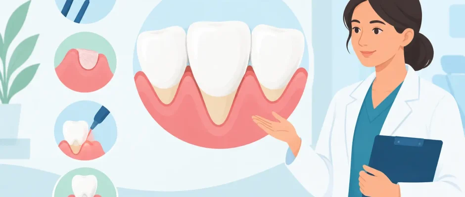
A tooth can look longer for reasons that are easy to miss at first: a notched area near the gumline, sudden sensitivity to cold, or a thin shadow where the root is becoming visible. This guide to gum recession treatment explains what can be treated conservatively, when surgery is justified, and why the correct diagnosis matters more than choosing a procedure quickly.
Gum recession is the apical movement of the gum margin, exposing part of the tooth root. It is not only an esthetic concern. Exposed root surfaces are more vulnerable to sensitivity, root decay, abrasion, and progressive tissue loss. In some cases, recession also signals inflammation or bone loss that needs attention before any cosmetic correction.
Guide to Gum Recession Treatment: Start With Diagnosis
The same visible recession can have very different causes. A patient may have healthy gums but naturally thin tissue and an aggressive brushing habit. Another may have recession caused by periodontitis, where inflammation has damaged the supporting bone and connective tissue around the tooth. Orthodontic tooth movement, crowding, an unfavorable bite, tobacco use, poorly positioned restorations, and piercings can also contribute.
This is why treatment should begin with a periodontal examination rather than a photograph alone. The clinician evaluates pocket depths, bleeding on probing, plaque control, the thickness and width of keratinized gum tissue, root exposure, tooth position, and the level of supporting bone. Digital imaging may be necessary when bone loss, previous treatment, or future implant planning is involved.
A key clinical question is whether the recession is stable. If inflammation is active or the cause remains unchanged, grafting the gum line may produce a disappointing or short-lived result. Treatment first removes the factors that are continuing to damage the tissue.
When recession needs prompt attention
Sensitivity is often the symptom that brings people to the dentist, but it is not the only reason to act. An exposed root can develop a cervical lesion, especially when acid exposure and strong brushing are combined. Recession on a tooth planned as an abutment for a bridge, veneer, or implant-supported restoration also requires careful planning.
Prompt assessment is particularly worthwhile when recession is progressing, bleeding occurs during brushing, a tooth feels mobile, or the gum line changes around an implant. Recession around an implant is not managed exactly like recession around a natural tooth. It may involve implant position, thin soft tissue, bone remodeling, or inflammation around the implant, and should be assessed with an implant-focused protocol.
First-Line Treatment: Control the Cause
Not every recession defect requires surgery. When tissue loss is mild and stable, the goal may be to protect the root and prevent further progression. A soft toothbrush, gentle technique, and a low-abrasion toothpaste can reduce mechanical trauma. The correct movement is small and controlled at the gum line, not forceful horizontal scrubbing.
Professional hygiene is equally important. Plaque that remains along the gum margin can maintain inflammation, while heavy calculus deposits cannot be safely removed by brushing at home. If periodontitis is present, periodontal therapy comes first. This may include professional debridement, hygiene instruction, reassessment, and a maintenance schedule tailored to the patient’s risk.
For sensitivity, desensitizing agents, fluoride varnish, or a protective bonded restoration may help. A small restoration can cover a non-carious cervical defect and reduce discomfort. However, it does not recreate lost gum tissue. When placed without considering the gum contour, it can make cleaning more difficult or compromise the appearance of later grafting.
If clenching, grinding, or a traumatic bite is contributing to overload, the plan may include occlusal adjustment only where clinically indicated, restorative correction, or a protective night guard. The aim is not to blame one factor for every recession, but to identify the combination relevant to that individual mouth.
When Gum Grafting Is the Better Option
Periodontal plastic surgery is considered when root coverage is predictably achievable and offers a meaningful benefit. Typical goals include reducing sensitivity, improving esthetics, increasing tissue thickness, protecting a vulnerable root surface, or preparing the area for restorative treatment.
The prognosis depends on several details: the height and width of recession, the amount of bone between adjacent teeth, the thickness of the gum tissue, tooth position, and smoking status. Complete root coverage is possible in many well-selected cases, especially when recession is limited and the tissue between teeth is intact. It cannot be promised in every defect. Clear expectations are part of safe treatment.
Connective tissue grafting
A connective tissue graft remains one of the most reliable approaches for many recession defects. A small amount of tissue is usually obtained from the palate and positioned beneath the gum around the affected tooth. The surgeon may use a tunnel technique or a coronally advanced flap to move and stabilize the gum over the exposed root.
Microsurgical instruments, fine sutures, and magnification support precise handling of delicate tissue. This matters because the blood supply to the graft and immobility during early healing strongly influence the result. The goal is not simply to cover a root, but to create a thicker, more stable gum contour that is easier to maintain over time.
The trade-off is that harvesting tissue from the palate creates a second healing area. Most patients manage this well with appropriate anesthesia, pain control, and postoperative instructions, but it should be discussed honestly before treatment. In selected cases, a collagen matrix may reduce the need for palatal harvesting. It can be useful, although the best option depends on the defect and the amount of tissue thickness required.
Regenerative materials and PRF
Biologic materials may be used as adjuncts in selected periodontal procedures. Platelet-rich fibrin, or PRF, is prepared from the patient’s own blood and can be placed to support wound healing. It does not replace meticulous surgical technique, correct graft selection, or careful aftercare. Its role is supportive rather than magical.
Similarly, enamel matrix derivatives and other regenerative materials may be considered when the root surface and supporting tissues meet specific clinical criteria. The decision should be based on anatomy and evidence, not on adding materials simply because they sound advanced.
What to Expect From Surgery and Recovery
Before surgery, the tooth and gums should be clean and stable. The procedure is usually performed under local anesthesia. For patients with substantial dental anxiety, additional comfort measures may be discussed as part of the treatment plan.
After grafting, the surgical area needs protection rather than vigorous cleaning. A soft diet, careful use of prescribed rinses, and avoidance of pulling the lip to inspect the site are usually advised during the first phase of healing. Brushing is temporarily modified around the graft, then gradually reintroduced according to the surgeon’s instructions. Sutures are commonly reviewed and removed within one to two weeks, depending on the technique.
Color and contour change gradually. Initial swelling, bruising, and an uneven appearance can be normal, particularly during the first days. Tissue maturation continues for months. Smoking and nicotine products can significantly impair healing and increase the risk of an incomplete result, so stopping before surgery and throughout recovery is strongly recommended.
Pain after soft-tissue surgery is often manageable, but it varies with the number of treated teeth, the use of a palatal donor site, and individual sensitivity. A detailed postoperative plan should state what discomfort is expected, how to manage it, and which symptoms require contacting the clinic, such as uncontrolled bleeding, increasing swelling after several days, fever, or severe pain.
Protecting the Result Over Time
Successful gum recession treatment is maintained at home and during professional follow-up. A graft can improve tissue thickness, but it cannot compensate indefinitely for forceful brushing, untreated inflammation, tobacco exposure, or a tooth that remains positioned outside the ideal bone envelope.
Regular periodontal reviews allow the clinician to compare measurements, monitor plaque control, and identify early changes before they become extensive. For patients considering veneers, crowns, orthodontics, or implants, the gum tissue should be part of the treatment discussion from the beginning, not an afterthought once the esthetic result is already compromised.
Recession is treatable, but the best result comes from a plan built around the cause, the anatomy, and the long-term function of the tooth. A careful examination can turn an alarming change in the mirror into a clear, measured path toward healthier and more stable gums.


