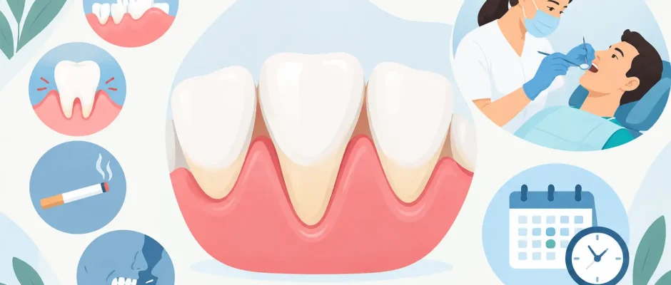
Gum Recession: Causes, Treatment, and Timing
A tooth can be perfectly healthy and still look longer than it used to. Often, the change is not in the tooth itself but in the gum around it. Gum recession occurs when the gum margin moves downward on a lower tooth or upward on an upper tooth, exposing part of the root surface. It may begin as a cosmetic concern, but sensitivity, root decay, and progressive loss of supporting tissue can follow.
The right response is not automatically surgery. Some recession is stable and can be monitored; other cases require early treatment to protect the tooth and restore a healthier gum contour. The difference becomes clear only after a careful periodontal examination.
What gum recession means for your teeth
The root is covered by cementum, not enamel. This surface is softer and less resistant to acids, abrasion, and bacterial plaque. Once exposed, it can become sensitive to cold air, cold drinks, or brushing. It also develops cavities more easily than the crown of a tooth.
Recession can affect one tooth or several teeth. A thin gum line around a lower front tooth may recede because of brushing trauma or tooth position. Recession around an implant, bridge, or crown can have different causes and needs separate evaluation. When recession is associated with periodontal disease, the concern is not only visible root exposure but also loss of the bone and attachment that stabilize the tooth.
A common misconception is that gums simply “grow back” after inflammation settles. Swelling may improve with hygiene and professional cleaning, making the gum line look healthier. But tissue that has truly receded does not usually return to its original position without a surgical procedure.
Why gum recession develops
Gum recession rarely has a single cause. The clinical goal is to identify the combination of factors that continues to pull the gum line away from the tooth.
Periodontal inflammation is one of the most significant causes. Plaque and tartar can trigger chronic inflammation, leading to gradual loss of gum attachment and bone. In this situation, a graft alone does not solve the problem. The inflammation must first be controlled through periodontal treatment and consistent home care.
Mechanical trauma is another frequent factor. Brushing with excessive pressure, using a hard-bristled brush, or scrubbing horizontally at the gum line can wear away gum tissue over time. This does not mean brushing less. It means using a soft brush, a gentle technique, and a toothbrush head that can reach the area without force.
Anatomy matters as well. Some people naturally have a thin gum phenotype, a narrow zone of attached gum, or a tooth positioned slightly outside the bony housing. Lower incisors and canines are particularly vulnerable. Orthodontic movement can be beneficial, but when a tooth is moved toward the outer boundary of the bone, the gum and bone support should be assessed before, during, and after treatment.
Other contributors include smoking, lip or tongue piercings, clenching with traumatic bite forces, poorly contoured restorations, and inflammation around teeth that are difficult to clean. In many cases, the recession is not a sign of poor hygiene or personal failure. It is a biological and anatomical problem that deserves a precise plan.
When gum recession needs treatment
Not every exposed root requires a gum graft. A stable recession defect without sensitivity, decay, inflammation, or cosmetic concern may be observed with periodic measurements and preventive care. Treatment becomes more strongly indicated when the recession is progressing, the root is sensitive, cavities recur, brushing causes discomfort, or the exposed area affects the smile.
The location matters. Recession around a lower front tooth may be monitored differently than recession around an upper canine visible in a broad smile. The amount of tissue between the tooth and the lip, the thickness of the gum, root prominence, and the presence of bone loss all influence the expected result.
A graft may also be recommended before restorative or orthodontic treatment if the gum tissue is too thin to tolerate future changes. This is especially relevant when long-term stability is the priority. Treating the tissue early can sometimes prevent a more complex problem later.
How the diagnosis is made
A proper assessment goes beyond looking in a mirror. The clinician measures the recession depth, probes around each tooth, evaluates bleeding and plaque, checks tooth mobility, and studies the bite. Digital photographs help document changes over time, while appropriate X-rays or three-dimensional imaging may be needed when bone loss, root anatomy, implant planning, or adjacent surgical treatment is involved.
The examination also determines whether root coverage is predictable. Complete coverage is often possible when there is no significant loss of bone and soft tissue between neighboring teeth. When that support has been lost, the objective may be to thicken and stabilize the gum rather than promise that every millimeter of exposed root can be covered.
This distinction is important. Careful treatment includes an honest discussion of what can be improved, what should be maintained, and what cannot be fully reversed.
Treatment for gum recession
The first stage is controlling the cause. For inflammation-related recession, this may include professional periodontal cleaning, treatment of deeper periodontal pockets when necessary, correction of home-care technique, and review of risk factors such as smoking. Sensitivity can often be reduced with desensitizing products or protective restorations placed conservatively on exposed root surfaces.
When soft tissue augmentation is indicated, microsurgical gum grafting can increase tissue thickness and, in selected cases, cover the exposed root. The most established approach often uses a small amount of connective tissue taken from the palate and positioned at the recession site. Depending on the anatomy, it may be combined with a coronally advanced flap that gently moves the existing gum toward the crown of the tooth.
Alternative materials may be appropriate in certain cases, particularly when several teeth require treatment or when minimizing a palatal donor site is a priority. The trade-off is that the choice of material and technique should be based on predictability, tissue quality, and the treatment goal, not convenience alone.
Microsurgical instruments, magnification, delicate sutures, and tension-free flap design are not cosmetic details. They help preserve blood supply and reduce trauma to the tissue during healing. In periodontal plastic surgery, small differences in tissue handling can affect comfort and the final contour of the gum line.
Recovery and long-term stability
After a gum graft, patients generally need a soft diet for a short period and should avoid pulling the lip to inspect the surgical site. Brushing is temporarily modified around the treated area, while antiseptic rinses and prescribed medications may be used according to the clinical protocol. Follow-up visits allow the surgeon to monitor healing and remove sutures when appropriate.
Most discomfort is manageable, but recovery depends on the procedure, the number of treated teeth, and whether tissue was harvested from the palate. Smoking and premature mechanical trauma can compromise healing. The tissue also continues to mature for weeks and months, so the early appearance is not the final result.
Long-term success depends on keeping inflammation under control and avoiding the original trigger. A soft toothbrush, gentle technique, regular periodontal maintenance, and protection from traumatic habits are part of treatment, not an afterthought. If clenching is contributing to overload, a night guard may be considered after the bite has been evaluated.
Recession around implants requires special attention
Gum recession around an implant is not identical to recession around a natural tooth. Implants lack the same connective tissue attachment and blood supply patterns found around teeth, so soft tissue management requires particular caution. Recession may expose an implant component, affect smile aesthetics, or make hygiene more difficult.
Prevention begins with three-dimensional implant planning. Implant position, available bone, soft tissue thickness, and the planned crown contour all influence the stability of the gum line. In complex cases, bone augmentation and soft tissue grafting may be part of planning rather than an attempt to correct a problem afterward. Digital planning and surgical guides can help place implants according to the restorative and biological requirements, not simply where bone appears most available.
A tooth that looks longer, feels sensitive, or bleeds during brushing is worth evaluating before the change becomes more difficult to treat. The most reassuring plan is a clear one: identify the cause, protect what is healthy, and use surgery only when it offers a meaningful, predictable benefit.


