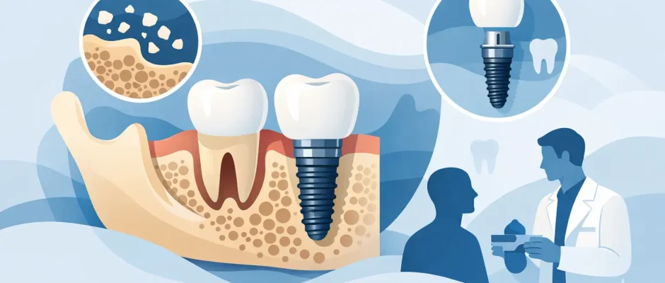
Bone Loss and Dental Implants Explained
A missing tooth does more than leave a gap in a smile. Once the root is gone, the jawbone in that area no longer receives normal chewing stimulation and can gradually shrink. This is why bone loss and dental implants must be planned together, not treated as separate problems. An implant needs a stable bone foundation, but reduced bone volume does not automatically mean that implant treatment is impossible.
The right solution depends on where the bone loss is located, how extensive it is, the condition of the gums, your general health, and the final restoration being planned. For some patients, an implant can be placed immediately after extraction. For others, grafting or a sinus lift creates the conditions for a safer, more predictable result.
Why bone loss happens after tooth loss
The jawbone is living tissue. It continually adapts to the forces placed on it. Natural tooth roots transfer chewing pressure into the bone. After an extraction, that stimulation disappears, and the ridge can become narrower and shorter over time. The most active changes often occur during the first months after tooth loss, although bone remodeling can continue for years.
Tooth loss is not the only cause. Long-standing gum disease can damage both the supporting bone and soft tissues around teeth. Infection at the root, trauma, poorly fitting removable dentures, and previous difficult extractions may also reduce the available bone. In the upper back jaw, the maxillary sinus can gradually expand downward after molars are lost, leaving little vertical bone for an implant.
This is why an implant consultation should not begin with the question, “Can an implant be placed?” A more useful question is, “What foundation is needed for the implant and crown to function well over time?”
Can dental implants be placed with bone loss?
Often, yes. The presence of bone loss changes the treatment plan, not necessarily the goal. Modern implantology offers several methods to rebuild or use the available bone strategically. The appropriate approach is chosen after a clinical examination and three-dimensional imaging, usually a cone beam CT scan.
A scan allows the surgeon to assess bone height, width, density, and the location of important structures such as the sinus cavity in the upper jaw or the mandibular nerve in the lower jaw. It also shows details that a standard two-dimensional X-ray can miss. This information is essential for selecting the implant position, length, diameter, and timing.
In selected cases, there is enough bone to place an implant without grafting, even if the ridge is not ideal. In other cases, placing an implant without creating adequate support may lead to an unfavorable angle, compromised appearance, difficult hygiene, or a higher risk of complications. The shortest path is not always the most durable one.
When immediate implant placement may be possible
An implant may sometimes be placed at the same appointment as tooth extraction. This can reduce the number of surgical stages and may help preserve the contours of the ridge. However, immediate placement is not appropriate simply because a patient wants treatment to move faster.
The socket must be free of uncontrolled infection, and there must be enough healthy bone to obtain primary stability for the implant. The gum tissue, bite, and position of the future crown also matter. In many cases, bone graft material and a membrane are placed around the implant to support the contour as healing occurs.
When these conditions are not present, a staged approach is often safer. The tooth is removed carefully, the site is preserved or grafted, and the implant is placed after healing. This is not a setback. It is a method of reducing risk and protecting the final result.
Treatment options for bone loss and dental implants
Socket preservation after extraction
When a tooth must be removed, socket preservation can help limit collapse of the bone ridge. After gentle extraction, graft material may be placed into the socket and covered with a membrane or healing material. The aim is not to stop all natural remodeling, which is impossible, but to preserve more volume for a future implant.
This procedure can be especially valuable in the visible front area, where both bone and gum contours affect the appearance of the final crown.
Guided bone regeneration
Guided bone regeneration, often called GBR, is used when there is not enough width or height for proper implant placement. Bone graft material is positioned in the deficient area and protected by a membrane. The membrane helps prevent soft tissue from growing into the space while the body forms new bone.
The graft may be synthetic, animal-derived, donor-derived, or taken from the patient, depending on the defect and treatment plan. There is no single best material for every clinical situation. The stability of the graft, blood supply, soft tissue closure, and surgical technique are just as important as the material itself.
Healing time varies. A smaller contour graft may heal alongside an implant, while a larger defect may require several months before implantation. The treatment sequence should be explained clearly before surgery, including what is planned at each stage and why.
Sinus lift for the upper back jaw
In the posterior upper jaw, bone loss is often combined with enlargement of the maxillary sinus. A sinus lift creates additional bone height by carefully lifting the sinus membrane and placing graft material beneath it.
Depending on the remaining native bone, the implant may be placed at the same time as the sinus lift or after the graft heals. The decision is based on stability and anatomy, not on a fixed protocol. Digital planning is particularly valuable here because the sinus anatomy can vary significantly from person to person.
Short implants and alternative positions
For certain patients, short implants may avoid the need for more extensive grafting. In full-arch cases, tilted implants and protocols such as All-on-4 can sometimes use available bone while avoiding anatomical structures. These options can be highly effective when indicated, but they are not interchangeable solutions for every missing tooth.
A treatment plan should compare the benefits and limits of grafting, shorter implants, alternative implant positions, and fixed versus removable restorations. The best choice is the one that supports function, hygiene, facial and gum esthetics, and long-term maintenance.
Why planning matters as much as the surgery
Bone augmentation and implant placement are precise surgical procedures. Predictability begins before the procedure with imaging, evaluation of your bite, periodontal assessment, and planning of the final tooth position. The implant should be placed for the crown, not simply wherever bone happens to be available.
Digital planning can combine CT data with an intraoral scan and the design of the future restoration. In appropriate cases, a surgical guide transfers that plan to the mouth and helps control implant position, depth, and angulation. A guide does not replace surgical judgment, but it adds accuracy and supports a more controlled procedure.
Biologic factors also matter. Active periodontal disease should be treated before implants are placed. Smoking, uncontrolled diabetes, some medications, teeth grinding, and inconsistent hygiene can affect healing and long-term implant health. These issues do not always exclude treatment, but they require honest discussion and risk management.
PRF, created from a small sample of the patient’s own blood, may be used in selected surgical protocols to support soft tissue healing and graft management. It is an adjunct, not a substitute for sound diagnosis, stable grafting, or careful postoperative care.
What recovery usually involves
Most patients are concerned less about the technical name of a procedure than about how it will feel. With effective local anesthesia and a calm, well-organized surgical protocol, implant and graft procedures are usually more manageable than expected. Swelling, mild bruising, and temporary discomfort are normal after more extensive grafting, particularly in the upper jaw.
Recovery instructions are tailored to the procedure. They may include medication, gentle oral hygiene, a soft diet for a short period, and avoiding smoking or activities that increase pressure in the sinus after a sinus lift. Follow-up visits are part of treatment, not an optional extra. They allow the surgeon to monitor healing, remove sutures when needed, and identify concerns early.
The implant does not become a finished tooth on the day it is placed. It needs time to integrate with the bone, followed by restoration with a custom crown, bridge, or full-arch prosthesis. The total timeline can range from a few months to longer in complex grafting cases. A longer plan can be worthwhile when it creates a more stable and maintainable result.
Questions worth asking at your consultation
A clear consultation should leave you with more than a price estimate. Ask what caused the bone loss, whether grafting is required, and what alternatives exist. Ask how the implant position will be planned, whether a surgical guide is appropriate, and how long each healing stage is expected to take.
You should also understand the risks of proceeding without grafting, the expected maintenance for the final restoration, and what happens if the site does not heal as planned. A responsible surgeon does not promise that every case will follow a perfect timetable. Instead, the goal is to use diagnostics, microsurgical technique, and thoughtful staging to make the outcome as predictable as possible.
Bone loss can make implant treatment more complex, but it does not have to make it uncertain. With accurate imaging, an individualized surgical plan, and careful follow-up, many patients can move from a compromised site to a stable, comfortable restoration built for everyday life.


