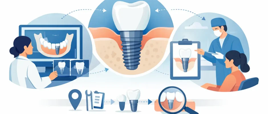
How Dental Implant Planning Works Step by Step
A dental implant is not selected from a catalog and placed wherever a tooth is missing. The surgical procedure may take a short time, but the quality of the result is largely decided beforehand. Understanding how dental implant planning works helps replace uncertainty with a clear sequence: diagnosis, risk assessment, restorative design, and a surgical plan tailored to your anatomy.
For some patients, this process leads to immediate implant placement on the day of extraction. For others, the safer route is to treat inflammation, rebuild bone, or allow tissues to heal first. A good plan does not force every case into one protocol. It identifies the protocol that gives the implant the best long-term conditions.
Implant Planning Starts With the Final Tooth
An implant is the foundation for a future crown, bridge, or full-arch restoration. That is why planning begins not only with the missing tooth but with the desired final result: where the new tooth should emerge, how it will meet the opposing teeth, how it supports the lips and smile, and how it can be cleaned.
This is called prosthetically driven planning. The surgeon evaluates the implant position in relation to the planned restoration, rather than simply placing it in the area with the most available bone. Bone matters greatly, but an implant placed too far toward the cheek, palate, tongue, or neighboring tooth can create esthetic, functional, or hygiene problems later.
For a single front tooth, millimeters can affect the gum line and symmetry of the smile. In the back of the mouth, planning focuses more heavily on chewing forces, access for cleaning, and critical anatomical structures. When several teeth are missing, the plan must also account for how forces will be distributed across the future bridge.
Clinical Examination: More Than Looking at the Gap
The first appointment includes a detailed examination of the teeth, gums, bite, and soft tissues. The goal is to understand why the tooth was lost and whether the same risk could affect an implant.
Periodontal disease, untreated cavities in adjacent teeth, clenching, uncontrolled diabetes, smoking, and inadequate oral hygiene do not automatically rule out implant treatment. They do, however, change the treatment sequence and maintenance plan. An implant cannot develop a cavity, but the surrounding bone and gum can become inflamed. Preventing peri-implant disease starts before surgery.
The examination also assesses the thickness and shape of the gums. In visible areas, thin gum tissue may increase the likelihood of recession or a gray shine-through effect around the crown. In selected cases, soft-tissue grafting is considered to create a more stable and natural-looking contour.
Your existing bite is equally important. If you grind your teeth or have an uneven bite, the implant restoration may need a different design, and a night guard may be recommended after treatment. Implants are firmly anchored in bone and do not have the same shock-absorbing ligament as natural teeth, so force control matters.
3D Imaging Shows What a Standard X-Ray Cannot
A panoramic X-ray provides a useful overview, but implant planning commonly requires cone-beam computed tomography, or CBCT. This 3D scan shows the height, width, and shape of the available bone in precise sections.
CBCT imaging also allows the surgeon to locate structures that must be protected. In the lower jaw, this includes the inferior alveolar nerve, which provides sensation to the lower lip and chin. In the upper posterior jaw, it includes the maxillary sinus. The roots of neighboring teeth, hidden bone defects, retained root fragments, and areas of infection can also become visible on a 3D scan.
The scan does not make treatment automatic. It is a diagnostic tool that must be interpreted alongside the clinical examination and the final restorative plan. For example, a scan may show enough bone height for an implant, but insufficient width for a stable and correctly positioned implant. That distinction changes the plan.
Measuring Bone and Deciding Whether Augmentation Is Needed
An implant needs enough healthy bone around it to achieve stability and support the surrounding tissues over time. The question is not simply, “Is there bone?” It is whether there is enough bone in the right position for the planned tooth.
After an extraction, bone naturally remodels. This is especially relevant when a tooth has been missing for years, after severe periodontal disease, or when infection has destroyed part of the socket. Depending on the defect, treatment may include socket preservation at the time of extraction, guided bone regeneration, a bone graft, or a sinus lift in the upper jaw.
These procedures can lengthen the overall treatment time, but they are not an unnecessary delay. In a deficient site, placing an implant without adequate support may compromise position, esthetics, or longevity. In other situations, particularly when the socket walls are intact and infection is controlled, immediate implant placement may be appropriate.
The choice depends on bone anatomy, gum condition, infection, implant stability, the position of the tooth, and the type of temporary restoration planned. A careful surgeon explains both the advantages and the limits of immediate treatment rather than presenting it as the right answer for every patient.
Digital Planning Connects the Scan to the Future Restoration
Digital implant planning software combines CBCT data with intraoral scans or detailed dental impressions. The result is a virtual model of the jaw, teeth, and planned crown or bridge.
The surgeon can then select the implant diameter, length, angulation, and depth before the procedure. This is particularly valuable near the sinus, nerve canal, or adjacent tooth roots. It is also useful in esthetic cases, where the implant must support a natural emergence profile through the gums.
For full-arch rehabilitation, including All-on-4 concepts, digital planning becomes even more important. The team must determine which teeth can be preserved, where implants can obtain stable anchorage, whether angled implants are beneficial, and how a fixed temporary bridge will be supported. The plan is based on available anatomy and restorative goals, not on a standard number of implants for every patient.
When a Surgical Guide Is Recommended
A surgical guide is a custom device produced from the digital plan. It rests on the teeth, gums, or bone and directs the implant drills according to the planned position, angle, and depth.
Guided surgery can improve precision and help make complex treatment more predictable, especially when space is limited or several implants are being placed. It may also support a minimally invasive approach in suitable cases. However, a guide is not a substitute for surgical judgment. The surgeon still needs to assess bone quality, tissue condition, and implant stability during the procedure.
Not every single implant requires a guide. In straightforward cases with clear anatomy, freehand placement by an experienced implant surgeon may be appropriate. The decision should reflect clinical benefit, not technology for its own sake.
The Surgical Plan Includes Comfort and Healing
Planning also covers what happens around the surgery itself. This includes local anesthesia, whether sedation is appropriate, the need for tooth extraction, the type of sutures, and the expected recovery period. Patients should know in advance whether they will leave with a temporary tooth, a removable appliance, or a healing cap.
In selected cases, platelet-rich fibrin, or PRF, may be used to support soft-tissue healing and grafting procedures. PRF is created from a small sample of the patient’s own blood and is used as part of a broader surgical protocol. It can be helpful, but it does not replace careful infection control, stable grafting, or proper postoperative care.
The plan should also include follow-up visits. These appointments allow the surgeon to check healing, remove sutures when needed, monitor graft maturation, and confirm that the implant is integrating properly before the final restoration is made.
How Dental Implant Planning Works in Complex Cases
Complex does not mean impossible. It means the plan needs more diagnostic detail and often more stages. A patient with severe bone loss may need guided bone regeneration before implantation. A patient with a failing tooth in the smile zone may need an atraumatic extraction, immediate implant consideration, connective-tissue management, and a carefully designed temporary crown.
Patients with multiple missing teeth may benefit from a staged approach that preserves remaining healthy teeth while restoring areas of greatest functional need first. Others are better served by a full-arch solution. The correct choice depends on prognosis, hygiene ability, anatomy, bite, expectations, and budget.
At Implantolog.co.il in Tel Aviv, treatment planning is approached as a surgical and restorative roadmap, with attention to 3D diagnostics, microsurgical principles, and the patient’s comfort throughout each stage.
A thoughtful plan gives you something more valuable than a date for surgery: it gives you a reasoned answer to why this implant, in this position, with this sequence of treatment, is the safest path toward a stable result.


