Bone Deficiency Implant Solutions That Preserve Options
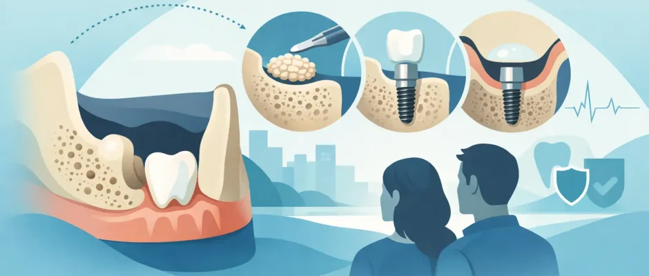
A missing tooth can begin changing the jawbone long before it becomes visible in the smile. After extraction, the bone that once supported the root gradually remodels. In some areas, especially the back of the upper jaw or a site missing a tooth for years, there may not be enough bone to place an implant in the ideal position. Bone deficiency implant solutions are designed to restore the conditions for stable, functional, and esthetic implant treatment – not simply to place an implant wherever bone happens to remain.
The right approach begins with a clear answer to one question: what is missing, and why? Bone height, width, density, the position of the sinus or mandibular nerve, gum thickness, bite forces, and the final crown design all affect the treatment plan. A careful plan can make even a complex case predictable. A rushed plan can create compromises that are difficult to correct later.
Why Jawbone Deficiency Matters for Implants
Dental implants need bone around them to distribute chewing forces and maintain long-term support. When bone is too narrow, too low, or irregularly shaped, placing an implant without preparation may lead to an unfavorable angle, exposed implant threads, insufficient stability, or an esthetic compromise in the visible zone.
Bone loss is not always severe, and it does not always require a major graft. The challenge is to determine whether the available bone can support an implant in the prosthetically correct position – the position that will allow the final tooth to look natural, be easy to clean, and function properly.
Several factors can contribute to bone deficiency. These include long-standing tooth loss, periodontal disease, infection around a failing tooth, trauma, previous extraction without socket preservation, removable dentures, and natural anatomy. In the upper posterior jaw, the maxillary sinus may limit available bone height. In the lower jaw, the location of the nerve canal must be respected at every stage.
Diagnosis Before Treatment: The Foundation of Bone Deficiency Implant Solutions
A panoramic image alone is often not enough for complex implant planning. Three-dimensional CBCT imaging shows the actual volume and shape of the bone, the proximity of anatomic structures, and possible inflammation that may need treatment first.
The surgical plan should also begin with the end result. Digital planning allows the implant position to be coordinated with the future crown or bridge rather than selected only according to the existing bone. When appropriate, a surgical guide can transfer that plan accurately during surgery. This is particularly valuable when available bone is limited, when several implants are planned, or when treatment involves the esthetic zone.
The consultation is also the time to review medical history, smoking status, oral hygiene, medications, gum condition, and bite. These details are not formalities. They influence healing, the choice of grafting material, timing of implant placement, and the long-term prognosis.
Treatment Options When Bone Volume Is Limited
There is no single procedure for every type of bone loss. The most conservative solution that provides stable conditions is usually preferred. In some cases, implant placement and bone augmentation can be performed together. In others, rebuilding the site first provides a safer and more predictable result.
Socket Preservation After Extraction
When a tooth must be removed, preserving the socket may reduce the collapse of the surrounding ridge. After atraumatic extraction, bone substitute material and, when indicated, a membrane may be placed in the socket. PRF, prepared from the patient’s own blood, can also be used as part of the healing protocol.
Socket preservation does not guarantee that no future grafting will be needed. It can, however, preserve valuable volume and may simplify later implant placement. This is why planning should begin before extraction whenever possible, especially in the front of the mouth.
Guided Bone Regeneration
Guided bone regeneration, or GBR, is commonly used when the ridge lacks width or has a localized defect. The surgeon places graft material around the deficient area and protects it with a barrier membrane. The membrane helps exclude fast-growing soft tissue cells while the bone regenerates underneath.
GBR can be performed at the time of implant placement if the implant has good primary stability and the defect is manageable. For larger deficiencies, a staged approach may be more appropriate: first regenerate bone, then place the implant after healing. This takes longer, but treatment time should not be the only measure of success. A stable foundation is often worth the additional months.
Bone Block Grafting
More substantial horizontal or vertical deficiencies may require a bone block graft. This approach is reserved for selected cases where particulate grafting alone is unlikely to provide adequate volume. It is technically demanding and requires meticulous stabilization and soft-tissue management.
The advantage is the ability to reconstruct a larger defect. The trade-off is a more involved procedure, a longer healing period, and, in some cases, a donor site if the patient’s own bone is used. A detailed discussion of alternatives is essential before choosing this route.
Sinus Lift in the Upper Jaw
In the posterior upper jaw, the sinus may leave only a few millimeters of bone beneath it. A sinus lift creates space below the sinus membrane so bone graft material can be placed and, eventually, support an implant.
A crestal, or internal, sinus lift may be suitable when some bone height is present and only limited elevation is required. A lateral window sinus lift is generally used for more pronounced deficiency. Whether an implant can be placed at the same appointment depends on the remaining bone, implant stability, and the overall surgical plan.
Short, Angled, or Alternative Implant Strategies
Not every patient with limited bone needs extensive augmentation. In selected cases, short implants, tilted implants, or strategically placed implants can avoid certain anatomic limitations. Full-arch protocols such as All-on-4 may use angled posterior implants to support a fixed bridge while avoiding sinus augmentation in appropriate candidates.
These strategies are not shortcuts for every patient. They depend on bone quality, jaw anatomy, the planned prosthesis, hygiene access, and bite forces. For a single missing tooth in the smile zone, for example, reconstructing the ridge may be preferable to accepting an implant position that compromises esthetics.
Immediate Implant Placement: When It Is and Is Not Appropriate
An implant can sometimes be placed immediately after extraction. This may shorten treatment and help preserve tissues, but it is not automatically the best choice. The presence of active infection, missing socket walls, inadequate primary stability, thin gum tissue, or an unfavorable implant position may make a staged approach safer.
When immediate placement is appropriate, precise extraction technique, three-dimensional implant positioning, grafting of the gap around the implant, and careful temporary restoration planning are critical. A temporary tooth should protect healing tissues without placing harmful pressure on the implant.
The goal is not to promise the fastest route. The goal is to select a protocol that protects the long-term result.
What Healing and Treatment Timing Can Look Like
Healing time varies with the procedure. A minor graft may heal alongside implant placement, while a larger augmentation may require several months before the implant is placed. After implant placement, osseointegration – the process by which bone bonds to the implant surface – also needs time before the final crown or bridge is delivered.
During this period, follow-up appointments matter. The surgical site is monitored, sutures are removed when needed, hygiene instructions are reinforced, and temporary restorations are adjusted. Pain is usually managed with local anesthesia during treatment and a clear postoperative medication plan. Swelling and discomfort can occur after grafting or sinus surgery, but they are typically temporary and should be addressed promptly if they become unusual or increase.
Smoking, uncontrolled diabetes, untreated periodontal inflammation, and inconsistent hygiene can raise the risk of complications. These factors do not always prevent implant treatment, but they may change the protocol and require closer management.
Choosing a Plan That Protects the Final Result
The best implant plan is not necessarily the least invasive on the day of surgery. It is the plan that preserves bone, respects anatomy, supports a cleansable restoration, and remains stable under function over time. In complex cases, experience in surgical dentistry, bone augmentation, and digital planning helps turn many difficult situations into structured, manageable treatment.
At Implantolog.co.il in Tel Aviv, treatment planning for bone deficiency is approached as a sequence of clinical decisions, not a standard package. CBCT-based diagnostics, guided surgery when indicated, microsurgical principles, PRF protocols, and careful follow-up are used to reduce uncertainty and protect healing.
If you have been told that there is not enough bone for implants, ask for a plan that explains the anatomy, the alternatives, the expected healing stages, and the reason each procedure is recommended. A well-planned reconstruction gives the future implant something essential: a foundation designed to last.


