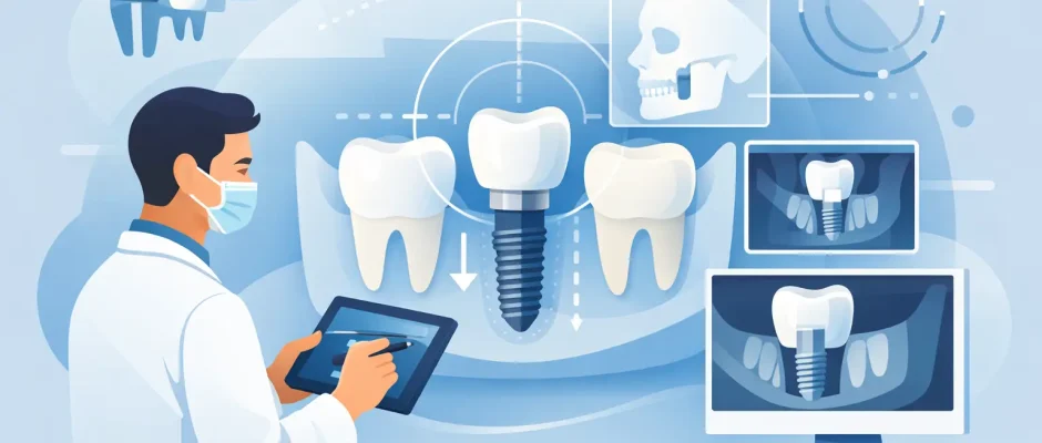
A dental implant is not simply placed where a tooth used to be. The real question is how dentists plan implant placement so the future crown has enough support, looks natural in the smile, avoids vital anatomy, and remains cleanable for years. That planning begins before surgery and, in complex cases, determines whether treatment is straightforward or requires preparation such as bone grafting, sinus elevation, or a staged approach.
For a patient, a detailed plan replaces uncertainty with a clear sequence: what can be done now, what must heal first, what the final restoration should look like, and why a particular protocol is being recommended.
Implant Planning Starts With the Final Tooth
Modern implant treatment is prosthetically driven. In practical terms, the surgeon first considers the tooth or teeth that need to be restored, then plans the implant position that can support that result. Placing an implant only where bone happens to be available can create compromises: a crown that is too wide or too narrow, poor emergence through the gum, an unfavorable bite, or a restoration that is difficult to clean.
The planned crown establishes the ideal position of the chewing surface, the line of the smile, contact with neighboring teeth, and the space needed for hygiene. From there, the clinician evaluates whether an implant can be placed in the correct three-dimensional position: front to back, side to side, and at the appropriate depth.
This is especially significant in the front of the mouth. A few millimeters can affect gum symmetry, the visible contour of the crown, and the support of the lip. In the back of the mouth, function and access for cleaning usually become the primary concerns, although esthetics still matter.
Clinical Examination: The Information a Scan Cannot Replace
A 3D scan is essential for implant planning, but it does not replace a careful clinical examination. The dentist evaluates the gums, tooth mobility, inflammation, bite pattern, existing crowns or bridges, and the amount of space between teeth. Photos, digital scans or impressions, and bite records help show how the teeth relate to the face and to each other.
The health of the surrounding tissues matters as much as the implant site. Untreated periodontal disease, active infection, uncontrolled diabetes, heavy smoking, teeth grinding, and poor plaque control can change the timing of treatment or the long-term risk profile. These factors do not automatically rule out implants, but they require an individualized plan and honest discussion.
A failing tooth also needs to be assessed carefully. Some teeth can be predictably saved with endodontic, periodontal, or restorative treatment. Others have fractures, severe bone loss, recurrent infection, or a poor long-term prognosis. Implant treatment should not be chosen by default. It should be chosen when it offers the better forecast for the specific clinical situation.
Planning the Extraction Site
When a tooth must be removed, the surgeon assesses the shape and condition of the socket. Is the outer bone wall intact? Is there acute infection? Are the gums thick enough to maintain a stable contour? Is there sufficient bone beyond the root tip to achieve primary stability?
If conditions are favorable, immediate implant placement may be possible. This means placing an implant during the same appointment as extraction. It can reduce treatment time and may help preserve tissue architecture, but it is not always the best choice. In the presence of significant infection, missing bone walls, insufficient stability, or an unfavorable implant position, socket preservation or delayed placement can be safer and more predictable.
Why CBCT Imaging Is Central to Implant Safety
Cone beam computed tomography, commonly called CBCT, provides a three-dimensional view of the jaw. Unlike a standard two-dimensional dental X-ray, it allows the surgeon to measure bone width and height, evaluate bone density patterns, and identify structures that must be protected.
In the lower jaw, the most important structure is often the inferior alveolar nerve, which provides sensation to the lower lip and chin. In the upper posterior jaw, planning must account for the maxillary sinus. The nasal cavity, adjacent tooth roots, and variations in anatomy also influence implant position and length.
CBCT planning answers practical questions: Is the bone wide enough for the planned implant? Is there enough vertical height? Is grafting needed? Can an implant be tilted safely to avoid an anatomic structure in a full-arch case? A scan does not make surgery automatic, but it gives the surgeon the information needed to reduce avoidable risk.
Deciding Whether Bone or Soft-Tissue Treatment Is Needed
After tooth loss, the jawbone gradually remodels. The longer a tooth has been missing, the more likely there is to be a deficiency in height or width. However, bone volume alone is not the only issue. The quality and shape of the bone, gum thickness, smile line, and planned restoration all matter.
For a narrow ridge, guided bone regeneration may be recommended. This can involve graft material and a protective membrane to rebuild volume before or during implant placement. In the upper back jaw, a sinus lift can create vertical bone where the sinus has expanded into the area of missing teeth. In selected cases, PRF may be used as part of a protocol to support soft-tissue healing and patient comfort.
There is a trade-off. A grafting procedure can add cost and healing time, yet it may permit an implant position that supports a stronger, more natural-looking, and easier-to-maintain restoration. In other cases, a shorter or angled implant, or a different restorative design, can avoid grafting without sacrificing predictability. The correct choice depends on the anatomy and the desired result, not on a single universal protocol.
Digital Planning and Surgical Guides
Digital planning combines CBCT data with an intraoral scan or a digital model of the teeth. Specialized software lets the surgical and restorative team view the proposed crown and implant together. The clinician can select implant diameter, length, depth, and angulation while verifying clearance from adjacent roots and anatomic structures.
When indicated, the digital plan can be transferred to a custom surgical guide. This guide rests on teeth, gums, or bone and directs the drilling sequence according to the approved position. Surgical guides are particularly valuable when precision is critical: in the esthetic zone, near vital anatomy, when multiple implants are being placed, or during full-arch rehabilitation such as All-on-4.
A guide is a precision tool, not a substitute for surgical judgment. It must fit accurately, and the surgeon must still evaluate the surgical site, bone quality, and implant stability during the procedure. If reality differs from the digital plan, an experienced clinician needs to recognize the issue and adapt safely.
Immediate Loading, Healing, and the Bite
Many patients ask whether they can leave with a tooth on the same day. Sometimes the answer is yes. Immediate provisionalization or immediate loading may be considered when the implant achieves sufficient primary stability, the bite can be controlled, and the patient can follow dietary and hygiene instructions.
It is not appropriate in every case. An implant placed into soft bone, an extensive grafting site, or a patient with a heavy grinding habit may need protected healing before it receives functional force. The goal is not the fastest protocol. The goal is stable integration of the implant with the bone and a restoration that can withstand normal chewing.
Bite planning is therefore part of implant planning. The final crown must distribute forces appropriately and avoid excessive sideways pressure. For patients with bruxism, a protective night guard may be recommended after the final restoration is delivered.
Planning Is Also a Conversation
A complete treatment plan should explain more than the surgical date. Patients should understand the number of stages, expected healing intervals, type of anesthesia, need for grafting, temporary tooth options, costs, and follow-up visits. They should also know what could change during treatment and why.
For patients traveling for care or coordinating treatment around work and family, this clarity is particularly valuable. At a surgical consultation, the objective is to create a plan that is clinically sound and realistic for the patient’s health, schedule, and expectations.
The best implant plan is often the one that feels least dramatic because every detail has been considered before surgery. A careful examination, 3D diagnostics, restorative planning, and precise surgical execution give the patient something more valuable than a quick procedure: a treatment path designed for safety, comfort, and a result that remains dependable over time.


