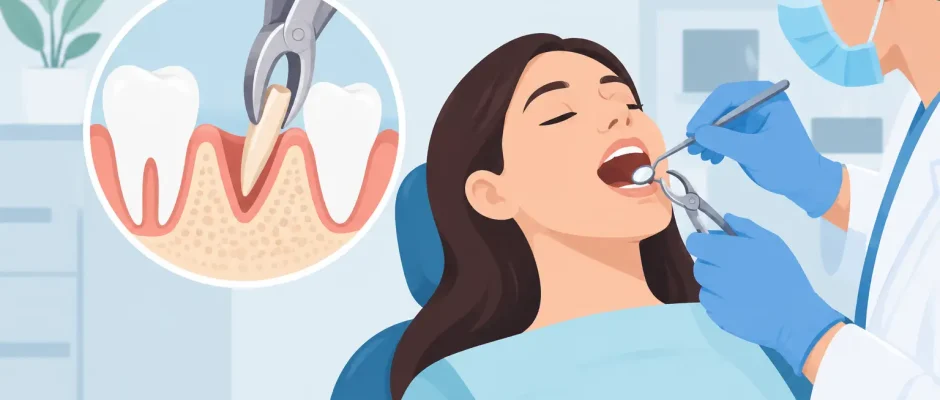
Retained Root Extraction Procedure: What to Expect
A tooth can break at the gum line with little warning: during chewing, beneath an old crown, after extensive decay, or while another dentist is attempting an extraction. What remains may be a retained root. A retained root extraction procedure is not simply a matter of “pulling out the rest.” The surgeon must determine where the root sits, how close it is to sensitive anatomy, whether inflammation is present, and how removal will affect the bone needed for future restoration.
For patients, the priority is straightforward: resolve pain or infection safely, preserve as much healthy tissue as possible, and leave a predictable path toward an implant, bridge, or other restoration when needed.
What Is a Retained Root?
A retained root is a portion of a tooth root that remains in the jaw after the crown has fractured or after a previous extraction. It may be visible above the gum, covered by soft tissue, or fully embedded in bone. Some roots are symptom-free for years. Others cause recurrent swelling, pain on biting, drainage, unpleasant taste, gum inflammation, or a chronic infection around the root tip.
The reason a root remains matters. Deep decay can weaken the tooth until the crown breaks off. Teeth that have undergone root canal treatment may become more brittle over time. In other situations, a root fragment is intentionally left during a difficult extraction because forcing it out would create a greater risk to the sinus, a nerve, or surrounding bone.
That distinction is clinically important. A retained root is not automatically an emergency, and extraction is not automatically the best first step. The correct decision follows examination and imaging, not an assumption based on an X-ray alone.
When a Retained Root Extraction Procedure Is Recommended
Removal is commonly advised when the root is associated with infection, pain, cystic changes, progressive bone loss, or repeated gum inflammation. Extraction may also be necessary when the root prevents implant placement, interferes with a planned denture, or lies beneath a failing restoration.
A root close to the surface can sometimes be removed through a relatively simple procedure. Deep roots, curved roots, roots fused to bone, and roots near the maxillary sinus or mandibular nerve require a more deliberate surgical approach. In the lower jaw, protecting the inferior alveolar nerve is particularly important because this nerve provides sensation to the lower lip and chin. In the upper back jaw, the concern may be the sinus floor.
There are cases in which monitoring is safer than removal. For example, a small, deeply positioned fragment without infection that lies immediately adjacent to a major nerve may be observed with periodic clinical and radiographic review. This is not “leaving a problem untreated.” It is a risk-benefit decision made when surgery may cause more harm than the retained fragment itself.
The Role of 3D Imaging
A panoramic X-ray provides useful initial information, but it is a two-dimensional image. When root position is uncertain or anatomy is complex, cone beam CT imaging can show the relationship between the retained root, surrounding bone, sinus, adjacent teeth, and nerve canals in three dimensions.
This information changes surgical planning. It helps the surgeon select the safest access point, estimate whether sectioning the root is needed, and decide whether bone grafting should be performed at the same visit. For a patient considering an implant, this level of planning is especially valuable because preservation of the extraction socket can influence the quality and volume of bone later available.
How the Procedure Is Performed
The retained root extraction procedure begins with local anesthesia. The goal is complete pain control, not simply tolerating the surgery. For patients with significant dental anxiety or for more involved procedures, additional sedation options may be discussed according to the clinical setting and medical history.
After the area is numb, the surgeon gently separates the gum tissue only when necessary to gain direct access. A small amount of bone may need to be carefully removed around the root. This is often preferable to applying excessive force, which can fracture surrounding bone or push a root fragment toward the sinus or nerve canal.
If the root is divided, it is done strategically. Sectioning allows individual pieces to be removed in a controlled direction rather than attempting to extract a large, resistant fragment at once. The socket is then cleaned of inflamed tissue or granulation tissue, inspected, and irrigated. If there is an acute infection, drainage and thorough debridement are often more important than antibiotics alone.
Depending on the condition of the socket and the future treatment plan, the surgeon may place grafting material and a membrane to preserve the ridge contour. Platelet-rich fibrin, or PRF, may also be used to support soft-tissue healing. When a small flap has been raised, sutures are placed and removed or allowed to dissolve according to the material used.
The details vary from case to case. A shallow retained root may take a short appointment. A root near the sinus, a deeply fractured molar, or an extraction site requiring bone preservation is a surgical procedure that deserves appropriate planning and time.
Can an Implant Be Placed at the Same Visit?
Sometimes, yes. Immediate implant placement may be considered when the infected tissue can be completely removed, the socket walls are sufficiently intact, and the implant can achieve stable fixation in healthy bone beyond the extraction site. It can reduce treatment time and help preserve tissue contours, but it is not the right choice simply because it is faster.
Delayed implant placement is often safer when there is extensive infection, insufficient bone, a large defect in the socket wall, or uncertainty about primary implant stability. In such cases, bone grafting and healing may create a more reliable foundation for the implant. A predictable result is more valuable than compressing every step into one visit.
For visible front teeth, the plan also considers gum thickness, smile line, and the ability to support natural-looking soft tissue. For back teeth, function, available bone height, and sinus anatomy may carry more weight. Digital planning and, when indicated, a surgical guide can help translate the diagnostic plan into accurate implant placement.
Recovery After Root Removal
Most patients experience mild to moderate soreness, swelling, and limited jaw opening for several days. The intensity depends on the location of the root, the amount of inflammation present before treatment, and the complexity of surgery. Discomfort is usually managed with the medication plan provided by the surgeon.
During the first 24 hours, protect the blood clot. Avoid vigorous rinsing, spitting, smoking, alcohol, and strenuous exercise. Choose soft, cool or lukewarm foods, and do not chew directly on the treated side. Cold packs applied externally in short intervals can help limit swelling on the first day.
Oral hygiene remains essential. Teeth away from the surgical area should be brushed normally. The extraction site should be cleaned only as instructed, often with gentle rinsing beginning the day after surgery. A follow-up visit allows the surgeon to assess healing, remove sutures when needed, and confirm that the next stage of treatment is proceeding as planned.
Contact the clinic promptly if pain becomes stronger after initially improving, swelling continues to increase after several days, persistent bleeding occurs, fever develops, or numbness remains or appears after the anesthesia should have worn off. These symptoms do not always signal a serious complication, but they should be assessed rather than managed at home.
Questions to Ask Before Surgery
A clear consultation should leave you knowing whether the root must be removed now, what imaging is needed, and whether the procedure has implications for future implant treatment. Ask about the root’s relationship to the sinus or nerve, the anticipated surgical approach, whether grafting is recommended, and the expected healing timeline.
It is also reasonable to ask what is included in the treatment plan: anesthesia, imaging, grafting materials, PRF, sutures, medications, and postoperative reviews. Clear answers reduce uncertainty and make it easier to compare treatment recommendations based on clinical value rather than price alone.
A retained root should be assessed before it becomes an urgent problem. With careful imaging, microsurgical technique, and a plan that accounts for future restoration, removal can be a controlled step toward comfort, healthy bone, and a stable long-term result.


