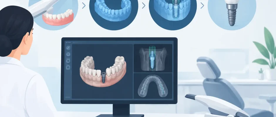
Digital Workflow in Implant Dentistry Explained
A missing tooth is not simply an empty space to fill. Its replacement must respect the available bone, the position of adjacent roots, the bite, gum contours, facial anatomy, and the future crown. A digital workflow in implant dentistry helps turn these variables into a clear treatment plan before surgery begins. For patients, that usually means fewer unknowns. For the surgeon, it means decisions can be made from measured clinical data rather than assumptions.
Digital tools do not replace surgical judgment or careful hands. They provide a more accurate way to collect information, evaluate risks, and transfer a plan to the operating field. This distinction matters most in complex cases: limited bone volume, proximity to the maxillary sinus or mandibular nerve, immediate implantation after extraction, and full-arch rehabilitation.
What a Digital Workflow in Implant Dentistry Includes
The workflow usually begins with a clinical examination and diagnostic imaging. A cone-beam CT scan provides a three-dimensional view of the jaw. Unlike a standard panoramic X-ray, it allows the surgeon to assess bone width and height, bone defects, root anatomy, the sinus floor in the upper jaw, and the course of the mandibular nerve in the lower jaw.
An intraoral scan records the teeth, gums, and bite without conventional impression material. The scan is then combined with the CBCT data in planning software. This creates a virtual model in which the planned implant can be positioned in relation to both the bone and the final prosthetic restoration.
That final point is essential. An implant should not be placed merely where bone happens to be available. It should be placed where it can support a functional, hygienic, and esthetic tooth. Sometimes the ideal crown position and the available bone do not align. The digital plan makes this discrepancy visible early, when there is time to consider bone grafting, sinus lift surgery, a different implant design, or an alternative restorative approach.
From Diagnostic Images to a Surgical Plan
Virtual planning allows the surgeon to select implant diameter, length, depth, and angulation while reviewing the surrounding anatomy in multiple views. The plan also considers the intended emergence profile of the crown and the space required between neighboring teeth or implants.
For a single tooth, this may sound straightforward, but millimeters matter. An implant placed too close to a natural tooth can compromise the supporting bone and soft tissue. An implant placed too far toward the cheek or palate may make the future crown difficult to clean or visually unnatural. In the posterior jaws, anatomy can limit the available space even further.
For full-arch treatment, such as an All-on-4 protocol, planning becomes even more interconnected. Implant positions must support a fixed bridge, distribute chewing forces appropriately, and avoid critical anatomical structures. The digital model also helps the surgical and restorative teams coordinate the provisional teeth that may be placed on the day of surgery when the clinical situation permits it.
When a Surgical Guide Is Useful
Once the virtual implant position is approved, a surgical guide can be manufactured. This is a custom device that rests on the teeth, gums, or bone and directs the drilling sequence according to the digital plan. Depending on the system and clinical objective, the guide may control the entry point alone or also guide the depth and angulation of implant placement.
Guided surgery is especially valuable when precision is critical: in narrow bone ridges, close to the sinus or nerve canal, in esthetically demanding areas, and in multiple-implant cases. It can also be helpful for immediate implantation, where the socket left after extraction may influence drill direction.
However, a guide is not an automatic guarantee of success. It depends on accurate imaging, a stable scan, proper guide seating, careful manufacturing, and the surgeon’s ability to recognize intraoperative changes. Severe infection, unexpected bone defects, limited mouth opening, or an unstable guide may require modification of the plan. Safe treatment always leaves room for clinical judgment.
Guided Does Not Always Mean Flapless
Some patients hear that a guided procedure is “flapless” and expect every implant to be placed without an incision. This is not always appropriate. A minimally invasive approach can reduce surgical trauma in selected cases, but it may not provide enough visibility when bone augmentation, removal of inflamed tissue, or contour correction is needed.
In a case with significant bone loss, opening the area may be the more responsible option. It enables direct assessment of the defect and precise placement of graft material and membranes during guided bone regeneration. The goal is not the smallest possible incision at any cost. The goal is stable bone, healthy soft tissue, and a predictable foundation for the implant.
Why the Workflow Can Improve Patient Comfort
Digital planning does not make surgery feel like a software demonstration. Local anesthesia, gentle tissue handling, sterile protocols, and postoperative care remain central to comfort and safety. Yet better preparation often makes the surgical phase more efficient because key decisions have been addressed beforehand.
Patients also benefit from seeing the plan. A three-dimensional image can make it easier to understand why an implant can be placed immediately after extraction in one situation but should be delayed in another. It can explain why sinus lift surgery is recommended, why a short implant may or may not be suitable, or why a failing tooth cannot always be saved predictably.
A clear plan is particularly helpful for people who are anxious about oral surgery. They should know what is included: diagnostic steps, anesthesia, the surgical procedure, possible grafting, sutures if required, follow-up visits, and the provisional restoration plan. Digital treatment planning supports this conversation, but it should never be used to promise an identical outcome for every patient.
Digital Planning in Complex Implant Cases
The value of digital diagnostics is often greatest when treatment is not routine. After long-term tooth loss, the jawbone may shrink in both height and width. In the upper posterior jaw, the maxillary sinus can limit implant length. In the lower jaw, the mandibular nerve must be protected. A CBCT-based plan helps identify these limitations before surgery rather than discovering them during it.
For immediate implant placement, the scan and virtual wax-up can help determine whether the remaining socket walls are sufficient and whether the implant can obtain primary stability in native bone. If these conditions are absent, a staged approach may be safer. Immediate treatment is a protocol, not a requirement.
When grafting is needed, the digital plan helps estimate the scale of reconstruction, although it cannot fully predict biological healing. Bone quality, smoking, periodontal inflammation, diabetes control, oral hygiene, and the ability to attend follow-up appointments can all affect the result. PRF may be used as an adjunct in selected surgical procedures, but it is not a substitute for sound grafting principles or meticulous postoperative care.
The Human Part of a Digital Treatment Plan
A high-quality scan is only the beginning. The result depends on how that information is interpreted and how carefully the plan is carried out. Implant dentistry requires an understanding of surgery, anatomy, healing biology, occlusion, and the restorative endpoint. It also requires the willingness to slow down when the safest plan is not the fastest one.
At Implantolog.co.il, digital diagnostics and surgical templates are used as part of a disciplined approach to implant treatment, including challenging cases involving bone deficiency and complex extractions. The treatment plan is individualized because a template cannot determine a patient’s health priorities, healing capacity, or expectations.
The most useful question is not whether treatment is digital or conventional. It is whether the diagnostic information is sufficient, the plan respects your anatomy, and the team has prepared for both the expected procedure and possible findings during surgery. When those conditions are met, technology becomes what it should be: a precise tool supporting calm, thoughtful care.


