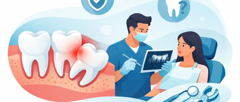
When Should Wisdom Teeth Be Removed Safely?
A wisdom tooth can remain quiet for years, then cause swelling, pain while swallowing, or pressure around a healthy second molar within days. So, when should wisdom teeth be removed? Not simply because they are called “wisdom teeth,” and not necessarily as soon as they appear. The right timing depends on their position, available space, condition of the surrounding bone and gums, and their effect on adjacent teeth.
For some patients, observation is appropriate. For others, delaying surgery can turn a relatively controlled procedure into treatment for infection, damage to the neighboring tooth, or a more difficult extraction. A clinical examination and appropriate imaging allow the surgeon to make this decision based on anatomy rather than guesswork.
When Should Wisdom Teeth Be Removed?
Removal is usually recommended when a third molar is causing disease, creating a significant future risk, or interfering with another planned treatment. The goal is not to remove teeth routinely. It is to preserve oral health while choosing the safest, most predictable approach.
A wisdom tooth often needs treatment when it is partially erupted and repeatedly inflames the gum around it. This condition, called pericoronitis, may cause tenderness, bad taste, limited mouth opening, facial swelling, or pain when chewing. Antibiotics can sometimes calm an acute infection, but they do not correct the food trap and bacterial pocket beneath the gum flap. If inflammation recurs, removal is often the durable solution.
Extraction may also be advised when decay affects the wisdom tooth or the tooth in front of it. A partially erupted lower wisdom tooth can be difficult to clean even for a highly motivated patient. Decay may progress unnoticed on the back surface of the second molar, where restoring the tooth can be challenging. In this situation, keeping the wisdom tooth may put a useful, fully functioning molar at risk.
Other common reasons include periodontal bone loss behind the second molar, a cyst or other abnormal lesion associated with the wisdom tooth, root resorption of the adjacent tooth, or pressure that complicates orthodontic or restorative planning. A tooth that is fully impacted may still require removal if imaging shows it is damaging nearby structures.
A Wisdom Tooth Does Not Need to Hurt to Need Evaluation
Pain gets attention, but it is not the only indication for surgery. Many impacted wisdom teeth are symptom-free because they are enclosed in bone. That does not automatically make them harmless.
On a panoramic X-ray or 3D scan, the surgeon may see that a lower wisdom tooth is leaning into the roots of the second molar, creating a deep periodontal defect, or placing that tooth at risk of resorption. In the upper jaw, a wisdom tooth may be close to the maxillary sinus, where repeated inflammation or decay can create more complex problems. These findings deserve a careful discussion even if the patient has no symptoms.
At the same time, an asymptomatic tooth with healthy surrounding tissues, no evidence of pathology, and a stable position may be monitored. This is especially relevant when removal carries higher surgical risk because of close proximity to the inferior alveolar nerve or the sinus. Follow-up is not neglect. It is an active plan that should include periodic clinical and radiographic review.
Age Matters, but It Is Not the Only Factor
Wisdom tooth surgery is often simpler in late adolescence or early adulthood because the roots may be less developed and the bone is generally more flexible. Healing can also be quicker. For that reason, when a problematic impaction is already evident, early planned treatment may be preferable to waiting for repeated infection or damage to occur.
However, adults in their 30s, 40s, 50s, and beyond can safely have wisdom teeth removed when there is a clear indication. The plan simply requires more attention to root anatomy, bone density, medical conditions, medications, and recovery expectations. Age alone is not a reason to remove or retain a tooth.
What Happens Before a Complex Extraction
A proper decision begins with diagnosis. The examination assesses gum inflammation, the ability to clean the area, the condition of the second molar, bite, and symptoms. A panoramic X-ray is often sufficient for an initial assessment. If a lower wisdom tooth appears close to the mandibular canal, or if the anatomy is unclear, a CBCT scan provides three-dimensional information.
This 3D assessment is particularly valuable for identifying the relationship between roots and the nerve that provides sensation to the lower lip and chin. It also helps evaluate bone coverage, root shape, and the most conservative surgical access. In the upper jaw, it shows the relationship to the sinus. Digital planning does not remove every risk, but it makes the procedure more controlled and allows the surgeon to explain the reasoning behind the chosen technique.
The discussion should include whether the tooth can be removed in one piece or requires sectioning, what type of anesthesia is appropriate, and what recovery may look like. For teeth with a high nerve-risk relationship, selected patients may be candidates for coronectomy – removing the crown while leaving stable roots in place. This is not suitable for every case, particularly where infection or root pathology is present, but it can reduce nerve-related risk in carefully selected situations.
When Waiting Can Create a Larger Problem
There is a difference between thoughtful monitoring and postponing care because a situation feels manageable. Recurrent swelling, drainage, fever, worsening pain, or difficulty opening the mouth should not be ignored. These can indicate an infection that needs prompt clinical assessment.
Waiting may also allow a cavity or periodontal defect to develop on the second molar. The wisdom tooth may be removable, but the damage to its neighbor can require root canal treatment, gum therapy, a crown, or, in advanced cases, extraction. Protecting the second molar is often one of the strongest reasons to address an unfavorable wisdom tooth before symptoms become severe.
Patients planning orthodontic treatment, implant rehabilitation, or extensive restorative work should also have wisdom teeth reviewed early. A third molar does not always interfere with these plans, but unresolved infection, poor access for hygiene, or a questionable adjacent tooth can affect the long-term result.
Recovery: What a Planned Procedure Should Include
Most patients can expect swelling and discomfort to peak during the first two to three days, then improve gradually. The exact recovery depends on whether the tooth was erupted, partially impacted, or deeply impacted in bone. Lower impacted wisdom teeth typically require a longer recovery than a straightforward upper tooth extraction.
A careful surgical protocol includes local anesthesia, gentle tissue handling, appropriate suturing when needed, and clear postoperative instructions. Pain control, cold compresses during the first day, soft foods, and avoiding smoking are practical parts of healing. Smoking and vaping can significantly increase the risk of dry socket and delayed healing.
PRF, prepared from a small sample of the patient’s blood, may be used in selected cases to support soft-tissue and bone healing. It is not a substitute for precise surgery or good postoperative care, but it can be a useful addition when the clinical situation calls for it.
Call Promptly if Recovery Changes Direction
Some discomfort is expected. Pain that becomes significantly worse after initial improvement, persistent bleeding, fever, spreading swelling, pus, rash, or numbness that does not follow the expected recovery pattern should be assessed promptly. Early review often prevents a minor postoperative issue from becoming more difficult to manage.
The Right Question Is Not “Do All Wisdom Teeth Need Removal?”
They do not. A fully erupted wisdom tooth that is healthy, cleanable, functional, and supported by healthy gums may be worth keeping. Conversely, a painless impacted tooth may still threaten the second molar or harbor disease that is visible only on imaging.
The best decision comes from balancing present symptoms against future risk, while accounting for the individual anatomy and surgical complexity. For patients with difficult impactions, a surgeon experienced in 3D diagnostics and complex extraction can turn an uncertain decision into a clear treatment plan with defined safety measures.
If a wisdom tooth has started to cause concern, do not wait for severe pain to decide. A focused examination can establish whether it should be monitored, treated conservatively, or removed at a time when the procedure and recovery are most predictable.


