Why Wisdom Teeth Become Impacted in Adults
A wisdom tooth can be fully healthy in structure yet still have no safe path into the mouth. That is why wisdom teeth become impacted: the tooth develops in a limited space at the very back of the jaw, where its position, angle, and surrounding bone may prevent normal eruption. An impacted wisdom tooth is not automatically an emergency, but it deserves a clear diagnosis rather than a decision based only on pain or an X-ray taken years ago.
What an impacted wisdom tooth means
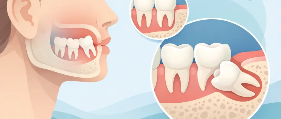
Wisdom teeth, also called third molars, are the last permanent teeth to develop. They are usually located behind the second molars and may begin erupting in the late teens or twenties. A tooth is considered impacted when it cannot erupt completely into a functional position because it is blocked by bone, gum tissue, or another tooth.
Some impacted teeth remain entirely within the jawbone. Others emerge only partly, leaving a flap of gum over part of the crown. This partially erupted position is particularly prone to repeated inflammation because food debris and bacteria can collect in an area that is difficult to clean.
Impaction is not the same as a tooth that simply erupts later than expected. Clinical examination and appropriate imaging help distinguish a delayed eruption from a tooth whose path is physically blocked.
Why wisdom teeth become impacted: the main causes
The most common reason is a mismatch between the size or shape of the jaw and the space required for the third molar. Human jaws vary considerably, and the available space behind the second molar can be limited even when the rest of the teeth look straight and well aligned.
Limited room behind the second molar
A wisdom tooth needs enough space not only for its crown but also for a proper eruption path. If the back portion of the lower jaw is short, or if the second molar occupies the available space, the third molar may remain trapped. In the upper jaw, limited space behind the second molar can create the same problem.
This is largely an anatomical and developmental issue. It is not caused by poor brushing, and it is not something a patient can prevent with exercises, supplements, or orthodontic retainers. Genetics influences jaw form, tooth size, and the direction in which a developing wisdom tooth forms.
An unfavorable angle of development
A wisdom tooth does not always grow upright. In the lower jaw, it may lean forward toward the second molar, backward toward the jawbone, sideways, or lie almost horizontally. A forward-leaning, or mesioangular, lower wisdom tooth is common. If its crown presses against the second molar, normal eruption may stop.
The angle matters because it changes both the likelihood of eruption and the treatment approach. A tooth that is nearly vertical and just beneath gum tissue may be relatively straightforward to remove. A deeply horizontal tooth surrounded by bone can require a more carefully planned surgical procedure.
Bone and soft-tissue obstruction
Sometimes the tooth has a favorable direction but remains covered by a thick layer of bone or gum tissue. During development, the root continues to form while the tooth remains enclosed. In some cases, the tooth is positioned unusually deep in the jaw, making spontaneous eruption unlikely even if some space appears present on a standard X-ray.
The timing of jaw growth and tooth development
Third molars develop later than the other permanent teeth. By the time they begin trying to erupt, the jaw has largely reached its mature dimensions and the adjacent teeth are already established. There is little ability for the mouth to create additional space at that stage.
This timing helps explain why wisdom teeth can be the only teeth affected. It also explains why crowding in the front teeth is not a reliable way to predict whether wisdom teeth will become impacted. A patient may have excellent alignment and still have deeply impacted third molars.
Why some impacted wisdom teeth cause problems
Many patients first learn about an impacted wisdom tooth after pain, swelling, or difficulty opening the mouth. These symptoms often arise from inflammation around a partially erupted lower wisdom tooth, known as pericoronitis. The gum tissue around the tooth can become tender, swollen, and difficult to clean. Symptoms may recur after temporary improvement.
An impacted tooth may also affect the second molar in front of it. When the two teeth are in close contact, plaque retention can contribute to decay or periodontal bone loss on the back surface of the second molar. This is clinically significant because the second molar is a key chewing tooth that should be protected whenever possible.
Less commonly, an impacted tooth can be associated with a cystic change around its crown or with resorption of the adjacent tooth root. These findings are not assumed from symptoms alone. They require careful evaluation of the images and the clinical situation.
It is also possible for an impacted tooth to remain symptom-free for years. Absence of pain does not prove that a tooth is risk-free, but it does mean that removal should not be presented as automatic. The decision depends on the tooth’s position, age of the patient, periodontal condition, ability to maintain hygiene, and evidence of current or likely damage.
How imaging guides a safe decision
A panoramic X-ray is often the first image used to assess wisdom teeth. It shows the general position of all four third molars, their relation to adjacent teeth, and the amount of available space. For many cases, this is enough to discuss whether monitoring or removal is reasonable.
For deeply impacted lower wisdom teeth, the key question is the relationship between the roots and the inferior alveolar nerve canal. This nerve provides sensation to the lower lip and chin. If a two-dimensional image suggests close proximity, a CBCT scan may be appropriate. CBCT provides a three-dimensional view and helps plan the access, bone removal, and tooth sectioning with greater precision.
In the upper jaw, imaging may also clarify the relationship to the maxillary sinus. The purpose is not to order advanced imaging routinely, but to use it when it changes the safety and predictability of the surgical plan.
When monitoring is reasonable and when removal is preferred
Monitoring can be appropriate for a fully impacted wisdom tooth that has no symptoms, no cystic changes, no damage to the second molar, and a position that makes future problems unlikely. Monitoring should be active, not forgotten: periodic clinical assessment and imaging at intervals recommended by the surgeon allow changes to be identified early.
Removal is more often recommended when there are repeated episodes of pericoronitis, decay that cannot be predictably restored, periodontal damage to the second molar, a cystic lesion, pain clearly linked to the tooth, or an unfavorable position with evidence of risk to the adjacent tooth. The best timing is individual. Earlier surgery can sometimes be technically simpler because roots may be less developed, while surgery later in life can still be performed safely with thorough planning and appropriate technique.
The trade-off is straightforward: surgery has temporary recovery and procedure-related risks, while retaining a problematic tooth may expose the patient to repeated infection or damage to a neighboring molar. A responsible recommendation accounts for both sides.
What careful surgical planning looks like
Surgical removal of an impacted wisdom tooth is not simply pulling a tooth. The plan may include local anesthesia, a carefully designed gum incision, conservative bone removal, and sectioning of the tooth into smaller parts when this reduces force on surrounding tissues. Sutures and postoperative follow-up are part of the treatment, not an afterthought.
For complex cases, the surgeon’s objective is to remove the tooth while preserving the adjacent second molar, surrounding bone, soft tissue, and nearby anatomical structures. Digital imaging, meticulous technique, and a clear explanation of expected recovery help replace uncertainty with a practical plan.
After surgery, swelling and discomfort usually peak during the first few days, then gradually improve. The exact recovery depends on the depth of impaction, the complexity of the procedure, the patient’s health, and adherence to postoperative instructions. Increasing pain after initial improvement, fever, persistent bleeding, worsening swelling, or altered sensation should be reported promptly.
An impacted wisdom tooth is an anatomical finding with a clinical context, not a diagnosis that demands the same answer for every patient. A focused examination and properly selected imaging can show whether the safest course is observation or planned removal – and give you a decision based on evidence rather than fear.


