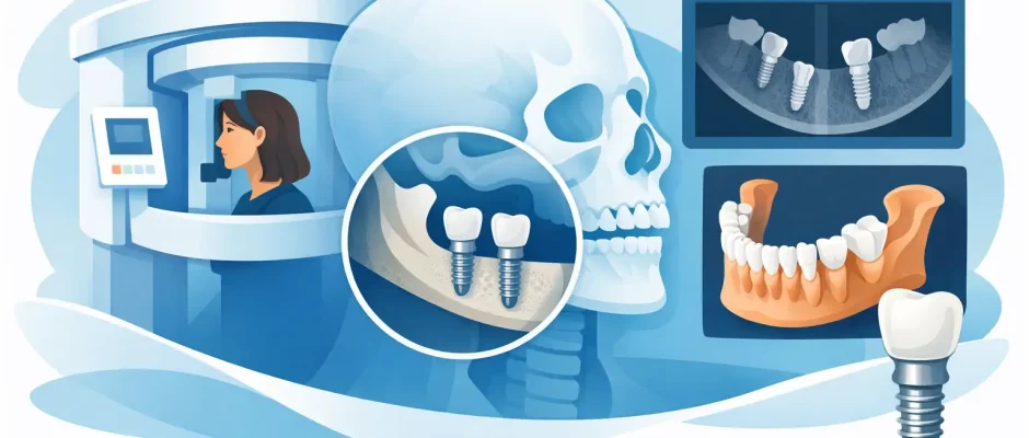
CT Scan for Dental Implants: What It Shows
A missing tooth can look like a straightforward problem, but the bone beneath it is never identical from one patient to another. A CT scan for dental implants provides the three-dimensional information needed to plan treatment around your individual anatomy, rather than making decisions from a two-dimensional image alone. For implant surgery, that difference can directly affect safety, predictability, and the long-term position of the final tooth.
In dentistry, patients often hear the term “CT scan” used for cone beam computed tomography, or CBCT. This imaging creates a detailed 3D model of the jaws. It allows the surgeon to assess not only whether an implant can be placed, but where it can be placed with the appropriate length, diameter, angle, and distance from important anatomical structures.
Why a CT Scan Is Used for Dental Implants
A standard dental X-ray is useful for many diagnostic tasks, including identifying infection, evaluating teeth, and reviewing the general level of bone. However, it compresses a three-dimensional structure into a flat image. It cannot reliably show the true width of the jawbone or the exact spatial relationship between an implant site and a nerve canal or sinus cavity.
A CT scan for dental implants shows the surgical site in thin cross-sections and in a 3D reconstruction. The clinician can measure available bone in millimeters, assess its contours, and plan the implant position before any incision is made.
This is particularly relevant in the posterior lower jaw, where the inferior alveolar nerve runs inside the bone. Injury to this nerve can lead to altered sensation in the lip or chin. In the upper posterior jaw, the maxillary sinuses can limit vertical bone height. A 3D scan helps identify these boundaries and determine whether implant placement is possible as planned or whether sinus lifting, bone grafting, or an alternative implant position should be considered.
The scan also reveals features that may not be visible on a conventional X-ray: a narrow ridge, an irregular bone defect after extraction, retained root fragments, an unrecognized cystic lesion, or the position of an impacted tooth. Finding these details before surgery is preferable to discovering them during surgery.
What the Surgeon Evaluates on the Scan
The image itself does not decide the treatment. Its value comes from careful clinical interpretation and integration with your oral examination, gum condition, bite, medical history, and restorative plan.
Bone volume and shape
An implant needs enough bone around it to achieve stable placement and to support healthy tissues over time. The scan provides measurements of bone height, width, and angulation. It also shows whether the ridge has resorbed after a tooth was lost.
Low bone volume does not automatically mean that implants are impossible. It may mean that the treatment plan should include guided bone regeneration, a sinus lift, a shorter or narrower implant in a suitable position, or a staged approach. The right choice depends on the specific defect and on the final restoration that needs to be supported.
Nerves, sinuses, and neighboring teeth
Safety margins are planned around anatomical structures. In the lower jaw, the nerve canal and, in some cases, the mental foramen must be located precisely. In the upper jaw, the floor and shape of the maxillary sinus are reviewed. The roots of neighboring teeth, as well as the position of existing implants, also influence where an implant can safely be placed.
A scan may show that a position which appears reasonable on a panoramic X-ray is too close to a nerve or sinus. Changing the plan before surgery is not a setback. It is part of responsible surgical planning.
Bone quality and potential inflammation
CBCT imaging can help assess the internal appearance of bone and detect areas of inflammation or pathology that require attention. Still, it is important to understand the limits of the technology: bone density values from dental CBCT are not always standardized in the same way as those from medical CT. The surgeon considers the scan alongside tactile feedback during preparation, implant stability measurements, and the overall clinical picture.
When a 3D Scan Is Especially Valuable
For many implant cases, 3D imaging is central to safe planning. It becomes particularly important when there is limited bone, when the implant site is near the sinus or a nerve canal, or when treatment involves more than one implant.
It is also valuable after a difficult extraction, in cases of previous implant failure, and when there has been periodontal bone loss or trauma. For full-arch rehabilitation, including All-on-4 concepts, the scan helps evaluate the entire jaw and plan implant angulation around anatomical limitations.
In immediate implant placement, where an implant may be placed at the time of tooth extraction, the scan helps assess the roots, the bony walls surrounding them, and the amount of bone available for primary stability. Immediate treatment can offer real advantages in selected cases, but it is not automatically the best protocol for every extraction. Active infection, thin facial bone, soft-tissue conditions, bite forces, and the patient’s ability to follow postoperative instructions all matter.
From Scan to Surgical Plan
The imaging appointment itself is usually quick and noninvasive. The patient stands or sits still while the scanner rotates around the head. No injections are generally needed for a dental CBCT scan. The process often takes less than a minute of actual scanning time.
Afterward, the scan is reviewed together with clinical records. The surgeon can virtually position the proposed implant, selecting its dimensions and angle while checking the planned distance from nerves, sinuses, and adjacent roots. The objective is not simply to place an implant where bone is available. It is to place it in a position that supports a functional, hygienic, and natural-looking crown or bridge.
This distinction matters. An implant placed too far toward the cheek, tongue, or palate may be stable initially but create compromises for the final restoration, gum contour, cleaning access, or load distribution. Prosthetic planning should guide surgical planning from the start.
When a surgical guide is recommended
In appropriate cases, digital planning can be transferred to a surgical guide. This is a custom device that rests on the teeth, gums, or bone and helps the surgeon follow the planned entry point, angle, and depth.
Guided surgery can improve precision and make the procedure more predictable, especially with multiple implants, limited bone, or full-arch treatment. It does not replace surgical judgment. The guide must fit accurately, the plan must account for the final restoration, and the surgeon must be prepared to adjust if clinical conditions differ from the digital model.
Radiation: A Balanced Perspective
Patients understandably ask whether a CT scan is safe. A dental CBCT scan uses ionizing radiation, so it should never be ordered routinely without a clinical reason. At the same time, the exposure is generally far lower than that of a conventional hospital CT scan, and modern dental systems allow the field of view to be limited to the area being examined.
The appropriate question is not whether radiation exposure is zero. It is whether the expected diagnostic benefit justifies the exposure. For implant surgery near a nerve, sinus, or thin bone ridge, a 3D scan can provide information that materially reduces uncertainty. The scan should be selected with the smallest suitable field of view and according to established radiation safety principles.
Pregnancy, recent imaging, and individual medical circumstances should be discussed before the scan. If you already have a recent, high-quality CBCT image that includes the necessary area, repeating it may not be required. The decision is individualized.
What a Scan Cannot Guarantee
A CT scan makes implant treatment more predictable, but it is not a guarantee of a complication-free result. Healing depends on many factors beyond bone anatomy, including smoking, uncontrolled diabetes, gum disease, oral hygiene, grinding, medications, and the quality of the final prosthetic restoration.
The scan is also a snapshot in time. If an extraction took place months earlier, bone may remodel. If treatment is delayed significantly after imaging, the surgeon may need updated information. Similarly, the digital plan must be confirmed during surgery, not followed mechanically.
For patients with advanced bone loss, the scan may show that a grafting procedure is needed before implants can be placed. While that can lengthen treatment, it may create a safer foundation and improve the position of the future teeth. A shorter plan is not always the better plan.
Preparing for Your Implant Consultation
Bring any previous X-rays, treatment records, and information about medications or health conditions. If you have had prior implants, extractions, sinus procedures, radiation therapy to the head or neck, or treatment with medications affecting bone metabolism, mention this at the first visit.
A careful consultation should leave you with more than a scan report. You should understand the condition of the bone, the proposed implant position, whether additional procedures are needed, and the alternatives if the preferred option is not appropriate. At Implantolog.co.il, digital imaging is used as part of a complete surgical plan, with attention to anatomy, restorative goals, and postoperative comfort.
The most useful scan is the one that turns uncertainty into a clear, clinically sound plan. Before moving forward with implant surgery, ask your surgeon to show you the key findings on the 3D image and explain how they shape the treatment recommended for you.


