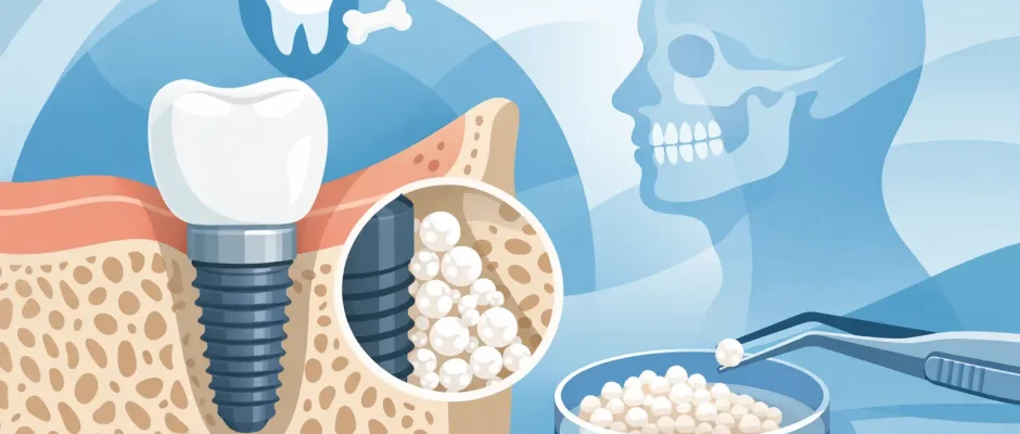
Top Reasons Dental Implants Need Bone Support
A dental implant may look like a replacement tooth, but its long-term success begins below the gumline. The top reasons implants need bone support are mechanical stability, healthy healing, natural esthetics, and protection of the surrounding tissues. Before placing an implant, the surgeon must determine not simply whether there is “some bone,” but whether there is enough bone in the right position and of sufficient quality for a safe, predictable result.
An implant is not cemented to neighboring teeth. It is a titanium fixture that becomes integrated with living bone. That biological connection is what allows the implant to support a crown, bridge, or full-arch restoration under daily chewing forces. When bone volume is limited, treatment may still be possible, but the plan should be based on three-dimensional diagnostics rather than assumptions.
Why dental implants need bone support
Bone holds the implant firmly in place
The first requirement is primary stability at the time of surgery. The implant must engage enough dense, healthy bone to remain stable while osseointegration takes place. During this healing phase, bone cells gradually form a direct attachment to the implant surface.
If the implant is placed into a narrow ridge, a shallow bone height, or bone weakened by long-term inflammation, it may not achieve sufficient initial fixation. Excess micromovement can interfere with healing. Instead of a stable bone-to-implant connection, fibrous tissue can form around the implant, increasing the risk of failure.
This does not mean that every patient needs bone grafting. Implant diameter, length, design, placement angle, and the quality of the remaining bone all matter. In selected cases, a shorter or narrower implant can be appropriate. In others, guided bone regeneration creates the foundation needed to place the implant in a safer position.
Bone distributes chewing forces
A natural tooth has a periodontal ligament that provides a small degree of cushioning. An implant does not. The forces from chewing pass from the crown through the implant directly into the surrounding bone.
Adequate bone around the implant helps distribute those forces. If support is insufficient, stress becomes concentrated in a smaller area. Over time, this can contribute to bone remodeling, loosening of prosthetic components, or overload of the implant itself, especially in patients who clench or grind their teeth.
The bite is therefore part of implant planning, not an afterthought. A single front-tooth implant, a back molar implant, and a full-arch All-on-4 restoration experience forces differently. The surgical and prosthetic plan must work together so that the final restoration does not ask too much of the available bone.
Bone determines where the new tooth can be placed
An implant should ideally be placed where the missing tooth was, not merely where bone happens to remain. This is particularly important in the visible front area of the upper jaw. If the ridge has shrunk after extraction, placing an implant too far toward the palate or too far toward the lip may create an unnatural crown shape and make hygiene more difficult.
Bone augmentation can allow the implant to be positioned according to the final restoration rather than as a compromise dictated by bone loss. Digital planning and surgical guides help translate that plan accurately into surgery. This improves control of implant depth, angulation, and distance from adjacent teeth and anatomical structures.
Sometimes a compromise remains necessary, even after grafting. A responsible treatment plan explains this clearly before surgery. The goal is not to promise a technically perfect result in every case, but to choose the option that offers the best balance of safety, durability, esthetics, treatment time, and cost.
Bone supports the gum contour and esthetic result
The contour of the gum is closely connected to the contour of the underlying bone. When bone is lost after an extraction, the gum often collapses with it. This can produce a visible indentation above an implant crown, uneven gum margins, or the appearance of a longer tooth.
In the smile zone, even a well-integrated implant can look disappointing if the surrounding hard and soft tissues have not been managed carefully. Bone grafting, soft-tissue grafting, atraumatic extraction techniques, and immediate implant protocols may be considered to preserve or rebuild the architecture around the tooth.
Immediate implant placement can be an excellent option in the right clinical situation, but it is not automatically the best choice. Active infection, thin facial bone, unfavorable socket anatomy, or inability to obtain stability may make a staged approach safer. The timing should follow the biology of the site, not a one-size-fits-all schedule.
What causes a lack of bone for implants?
Bone loss is common after tooth loss because the jaw no longer receives normal stimulation from the tooth root. The greatest changes often occur during the first months after extraction, but resorption can continue for years. The longer a tooth has been missing, the more likely it is that the ridge has become narrower and lower.
Other causes include periodontal disease, infection around a tooth root, trauma, removable dentures that place pressure on the ridge, and previous unsuccessful implant treatment. In the upper back jaw, the maxillary sinus can gradually expand downward after molars are lost, reducing the available vertical bone height. In the lower jaw, the surgeon must also maintain a safe distance from the nerve canal.
A conventional dental X-ray is useful, but it cannot show every dimension of the bone. Cone beam CT imaging provides a three-dimensional view of bone width, height, density patterns, sinus anatomy, and nerve location. This information is central to planning complex cases safely.
When bone grafting is the right option
Bone grafting is not a procedure performed simply because more bone would be convenient. It is recommended when the existing bone cannot reliably support an implant in its ideal restorative position or when insufficient bone would compromise safety or esthetics.
For a localized defect, guided bone regeneration may be used. The surgeon places grafting material in the deficient area and protects it with a membrane while the body forms new bone. In other cases, a block graft, ridge-splitting technique, or sinus lift may be more suitable. A sinus lift increases available bone in the posterior upper jaw by carefully elevating the sinus membrane and creating space for graft material.
Healing time depends on the procedure, the size of the defect, the patient’s biology, and whether an implant can be placed simultaneously. Some grafts allow implant placement in the same procedure; others require several months of healing before placement. Platelet-rich fibrin, or PRF, may be used as part of the protocol to support soft-tissue healing and regeneration, although it does not replace a properly planned graft when substantial volume is needed.
Smoking, uncontrolled diabetes, active periodontal disease, and poor oral hygiene can affect healing and should be addressed before or during treatment. These factors do not always rule out implant therapy, but they may change the timing, technique, and expected risk profile.
Alternatives when bone is limited
A bone graft is often valuable, but it is not the only path. Depending on the anatomy, an experienced implant surgeon may consider short implants, angled implants, implants placed in denser available bone, or a prosthetic solution that reduces the number of implants required. For a full arch, protocols such as All-on-4 can sometimes avoid extensive grafting by using strategically angled posterior implants.
These alternatives have trade-offs. Avoiding grafting may shorten treatment, but it can limit implant position or increase the demands on bite design. More extensive reconstruction may create an ideal foundation, but it adds surgery, healing time, and cost. The correct choice depends on the patient’s anatomy, medical history, expectations, and priorities.
A predictable implant result is not defined by placing an implant as quickly as possible. It is defined by placing it where it can heal securely, function comfortably, and support a restoration that can be maintained for years. A careful consultation with clinical examination, CBCT evaluation, and a clear written plan turns bone support from an abstract concern into a practical decision about the safest route to restoring your smile.


