Is CBCT Safe? What Dental Patients Should Know
A 3D dental scan can make a complex treatment plan feel more concrete, but it also raises a fair question: is CBCT safe? For most patients, cone beam computed tomography, or CBCT, is safe when it is prescribed for a clear clinical reason and performed with the smallest practical scan area and exposure settings. It should not be taken routinely simply because 3D images are available.
In implant dentistry and oral surgery, the value of CBCT is not the image itself. It is the ability to see anatomy that cannot be assessed reliably on a standard 2D X-ray: bone width and volume, the course of a nerve, the floor of the maxillary sinus, root position, and hidden pathology. That information can prevent avoidable surgical complications and support a more predictable plan.
What a CBCT Scan Actually Shows
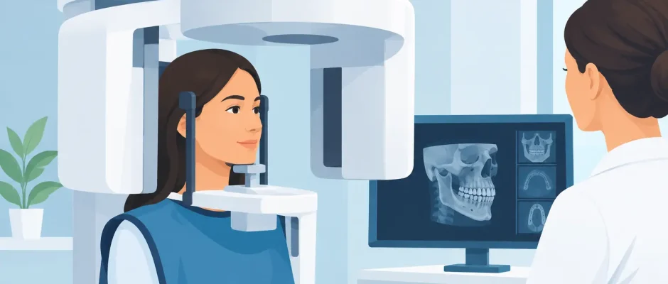
A CBCT scanner rotates around the patient’s head and creates a three-dimensional image of the teeth, jaws, and surrounding structures. The scan itself is quick. You remain still while the unit rotates, usually without entering an enclosed tunnel, and there is no pain or recovery time.
Traditional dental X-rays flatten a three-dimensional structure into a two-dimensional picture. They remain very useful for routine examinations, cavities, periodontal evaluation, and follow-up. However, overlapping structures can conceal clinically relevant details. This limitation matters when planning surgery near the mandibular nerve, evaluating an impacted wisdom tooth, or selecting the position, length, and angulation of a dental implant.
CBCT should also not be confused with a conventional medical CT scan. Both produce cross-sectional images, but dental CBCT is designed specifically for the maxillofacial region and can use a smaller field of view for a focused dental question.
Is CBCT Safe for Dental Implants and Surgery?
CBCT uses ionizing radiation, so the honest answer is not that it carries zero risk. Any medical imaging that uses X-rays should be justified. The relevant question is whether the expected diagnostic benefit outweighs the radiation exposure for your individual case.
For implant planning, that benefit is often substantial. A CBCT scan can show whether there is enough bone for an implant, where the inferior alveolar nerve lies in the lower jaw, and how close an implant site is to the sinus in the upper jaw. It can also identify retained roots, inflammation, cystic changes, or bone defects that could change the treatment sequence.
In other words, the scan may help the surgeon avoid placing an implant in an unsafe position, perforating the sinus membrane, or approaching a nerve too closely. For a patient with limited bone volume, a complex extraction, or planned bone grafting, a 3D image can be the difference between an assumption and a measured surgical plan.
The same principle applies to impacted wisdom teeth. When the roots appear close to the lower jaw nerve on a panoramic image, CBCT may clarify the actual relationship. In some cases, that information changes the operative technique or supports a more conservative approach.
Radiation Exposure Depends on How the Scan Is Taken
There is no single radiation dose for every CBCT examination. Exposure varies according to the machine, the field of view, image resolution, patient size, and settings selected by the clinic. A small scan focused on one implant site is fundamentally different from a large-volume scan of both jaws.
This is why responsible imaging begins before the scan. The clinician should define the clinical question and choose the smallest field of view that answers it. High-resolution settings may be useful for a specific diagnostic task, but they are not automatically necessary for every patient.
The guiding principle is often called ALARA, meaning radiation exposure should be “as low as reasonably achievable.” In dental imaging, many clinicians also use the concept of ALADA: as low as diagnostically acceptable. The distinction is meaningful. The goal is not simply the lowest possible dose, but the lowest dose that still provides an image accurate enough to guide treatment safely.
A properly selected CBCT scan is therefore a targeted diagnostic procedure, not a routine extra. If a conventional X-ray answers the clinical question, CBCT may not be needed. If 2D imaging leaves uncertainty that could affect surgery, CBCT may be the safer choice overall.
When a CBCT Scan Is Usually Justified
CBCT is particularly valuable when the treatment plan depends on precise three-dimensional anatomy. Common examples include implant placement, immediate implant placement after extraction, sinus lift procedures, guided bone regeneration, assessment of severe bone loss, and full-arch treatment such as All-on-4.
It may also be indicated for difficult extractions, impacted teeth, root fractures, resorption, suspected perforations, persistent infection after root canal treatment, and planning microsurgical procedures such as apicoectomy. In these situations, a small anatomical detail can influence the treatment decision.
For guided implant surgery, CBCT data can be combined with a digital scan of the teeth. This allows the restorative and surgical plan to be coordinated before the procedure. A surgical guide can then help transfer the approved implant position to the mouth. The guide does not replace surgical judgment, but it adds another level of control when used for the right indication.
When It May Not Be Necessary
A CBCT scan is not automatically required before every extraction, implant consultation, or dental complaint. Repeating a scan without a new clinical reason is usually not appropriate, especially if a recent diagnostic-quality scan is already available.
Routine checkups, uncomplicated cavities, and many straightforward treatments can be evaluated with lower-dose conventional dental radiographs. An image should be taken because it can change diagnosis or treatment, not because it is technologically impressive.
Patients should also understand that a 3D scan cannot compensate for poor planning. Its usefulness depends on careful interpretation, an accurate clinical examination, and a surgeon who knows which findings actually matter. Technology improves safety when it is integrated into a disciplined protocol.
Pregnancy, Children, and Other Individual Considerations
If you are pregnant or think you may be pregnant, tell the dentist or surgeon before any X-ray examination. Dental imaging is not necessarily prohibited during pregnancy when there is a genuine clinical need, but elective scans are commonly postponed when this can be done without compromising care. The decision should be individualized and based on urgency.
Children and adolescents deserve special consideration because they are more sensitive to radiation and have a longer lifetime in which radiation-related effects could theoretically occur. For younger patients, strict justification, a limited field of view, and child-appropriate exposure settings are especially important.
Prior imaging also matters. Bring previous panoramic X-rays, CBCT scans, and medical reports to your consultation when possible. Reviewing existing records may prevent unnecessary repeat imaging and helps create a more complete clinical picture.
Questions Worth Asking Before Your Scan
You do not need to become an imaging expert, but you should feel comfortable asking why a scan is recommended. A clear explanation usually includes the clinical question, the area being scanned, and how the result will influence the treatment plan.
Useful questions include: What information will CBCT provide that a regular X-ray cannot? Can the scan be limited to the specific area of concern? Will the result affect whether I need an implant, bone graft, sinus lift, or a different surgical approach? If you have a recent scan, ask whether it can be used rather than repeated.
A careful consultation should turn the scan into part of a visible plan: diagnosis, options, surgical sequence, expected healing, and follow-up. This is particularly reassuring for patients facing complex implant rehabilitation or surgery after years of missing teeth.
The Safer Choice Is an Informed One
CBCT is neither something to fear automatically nor something to request indiscriminately. Used selectively, with an appropriate field of view and a real diagnostic purpose, it provides information that can make oral surgery and implant treatment more precise.
Before proceeding, ask your surgeon to show you what the scan will help evaluate in your specific case. When you understand the anatomy, the alternatives, and the reason behind each step, a complex treatment plan becomes much easier to approach with confidence.


