How Much Bone Is Needed for Dental Implants?
The question, “how much bone is needed for dental implants,” has no single millimeter answer. A dental implant needs enough healthy bone around it to remain stable under chewing forces, but the right plan depends on the implant site, bone quality, neighboring anatomy, bite, and the restoration that will be placed on top. A three-dimensional CBCT scan and a prosthetically driven plan are more useful than a general rule heard online.
For many patients, reduced bone volume does not mean implants are impossible. It means the surgical approach must be selected carefully – whether that involves a narrower or shorter implant, bone grafting, a sinus lift, or a full-arch protocol that uses available bone strategically.
How Much Bone Is Needed for Dental Implants?
Implant planning considers bone height, bone width, and bone quality. The goal is not simply to place an implant where bone exists. The goal is to place it in a position that supports a functional, hygienic, and natural-looking tooth while respecting the sinus, nerves, roots of adjacent teeth, and soft tissues.
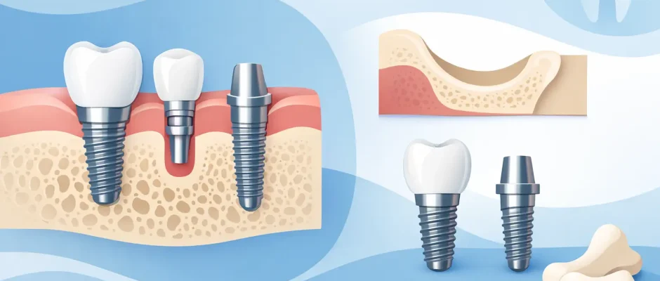
As a broad clinical reference, a conventional implant often benefits from approximately 8 to 10 mm of available bone height and about 6 mm of bone width. These figures are not a universal requirement. Modern implant systems include shorter and narrower designs, and some cases can be treated safely with less native bone. At the same time, using an implant that is too narrow, too short, or poorly positioned just to avoid grafting may create compromises in long-term stability, esthetics, or load distribution.
The most relevant measurement is not the amount of bone on a panoramic X-ray. It is the three-dimensional volume of bone in the exact intended implant position.
Bone height: the vertical dimension
Bone height is measured from the crest of the jaw to an important anatomical boundary. In the upper back jaw, that boundary is usually the maxillary sinus. In the lower back jaw, it is the mandibular canal, which contains the inferior alveolar nerve. A safety margin must be maintained around these structures.
Loss of a back upper tooth often leads to two changes at once: the ridge becomes lower after extraction, and the sinus may expand downward. This is why the posterior maxilla is one of the most common areas where a sinus lift is considered. In the lower jaw, the available height may be limited by the position of the nerve canal, requiring precise digital planning.
Bone width: often the hidden limitation
A ridge may look tall enough on a standard two-dimensional image but be too thin for ideal implant placement. An implant requires bone on both the cheek side and tongue or palate side. If the facial bone is extremely thin, it is more vulnerable to resorption and can affect gum contours over time, especially in the visible smile zone.
Width is particularly important for front teeth. A successful result there is not only about integration of the implant. It is also about supporting the gum line, papillae between teeth, and the emergence profile of the future crown.
Bone quality: density changes the plan
Not all bone holds an implant the same way. Dense lower-jaw bone may provide strong initial fixation even with limited dimensions. Softer bone in the upper posterior jaw can require a different drilling protocol, implant design, or healing period. Bone quality is assessed on CBCT imaging and confirmed during surgery.
Initial implant stability matters most when immediate loading or immediate placement after extraction is being considered. It is one reason why two patients with similar measurements may receive different recommendations.
Why Bone Is Lost After Tooth Extraction
The jawbone is maintained partly by the forces transmitted through the tooth root. After a tooth is removed, the ridge naturally remodels. The greatest dimensional reduction often occurs during the first months after extraction, particularly on the facial side of the ridge.
Bone loss may be more extensive when a tooth has been missing for years, when there was a chronic infection, or when periodontal disease has affected the supporting tissues. Trauma, poorly fitting dentures, sinus expansion, and long-term pressure from an unstable bridge can also contribute.
This does not mean every extraction should automatically be followed by a graft. The decision depends on the condition of the socket, the timing of implant placement, and the intended final tooth position. When appropriate, immediate implant placement and socket preservation can reduce the number of surgical stages, but neither approach is suitable for every site.
When Bone Grafting Is Recommended
Bone grafting is recommended when the existing ridge cannot securely support an implant in its ideal restorative position or when insufficient bone would compromise the surrounding soft tissue. The procedure is designed to rebuild or preserve volume, not to make an image look better.
For a narrow ridge, guided bone regeneration may be performed using graft material and a protective membrane. Depending on the defect, the surgeon may also use autogenous bone, meaning bone from the patient, or a combination of materials. PRF, made from the patient’s own blood, may be used to support wound healing and soft-tissue management in selected cases.
A sinus lift addresses limited bone height in the upper back jaw. During this procedure, the sinus membrane is gently elevated and graft material may be placed beneath it to create adequate vertical space. In some situations, an implant can be placed at the same appointment. In others, staged healing provides a safer and more predictable path.
Grafting adds time and cost, so it should never be presented as a routine upsell. However, skipping a necessary graft can lead to an implant positioned too far toward the palate, too far to one side, or outside the bone envelope. That may complicate the crown design, hygiene, appearance, and long-term maintenance.
Can Implants Be Placed Without a Bone Graft?
Often, yes. The decision is based on risk and prosthetic requirements rather than a simple yes-or-no measurement. Short implants may be appropriate in areas with reduced height. Narrow implants may work in selected thin ridges. In the upper posterior jaw, techniques such as tilted implants can sometimes avoid sinus grafting in full-arch rehabilitation.
For patients missing all teeth in one jaw, All-on-4 or related full-arch concepts may use the denser available bone in the front part of the jaw. Angled posterior implants can help avoid the sinus in the upper jaw or the nerve canal in the lower jaw. This approach can be highly effective when properly indicated, but it is not interchangeable with replacing a single tooth. The bite, bone distribution, prosthetic design, hygiene access, and medical history all matter.
There is also a trade-off between fewer surgical stages and ideal tissue reconstruction. A graft-free solution may shorten treatment in the right case. In another case, rebuilding the ridge first may provide a better esthetic and functional result for decades.
How a Precise Plan Is Created
A reliable implant plan begins with examination of the gums, remaining teeth, bite, and medical history. A CBCT scan provides three-dimensional information about bone volume and nearby anatomical structures. Digital planning then determines implant diameter, length, angle, depth, and relationship to the future crown.
The crown should guide the implant position, not the other way around. This is called prosthetically driven planning. In complex or high-precision cases, a surgical guide can transfer the digital plan to the clinical setting and improve control of position and angulation.
The plan also includes timing. An implant may be placed immediately after extraction, after several weeks of soft-tissue healing, or after bone augmentation has matured. The correct timing is the one that balances infection control, stability, bone preservation, and the patient’s goals.
Questions Worth Asking at Your Consultation
A clear consultation should explain how much bone is present in the planned implant site, where the anatomical limitations are, and whether the implant can be placed in the ideal restorative position. You should also understand whether grafting is necessary, optional, or avoidable with a different implant strategy.
Ask how the final crown or bridge will be planned, what healing time is expected, and what alternatives exist if you prefer to avoid additional surgery. For patients with gum disease, smoking, uncontrolled diabetes, teeth grinding, or a history of failed implants, risk management should be discussed openly before treatment begins.
At Implantolog.co.il in Tel Aviv, complex implant cases are planned with CBCT-based diagnostics, digital workflows, and microsurgical principles when they are clinically indicated. The purpose is not to offer the same procedure to every patient, but to choose the least invasive approach that still protects the result.
The amount of bone is only the starting point. A well-planned implant treatment considers the bone you have, the tooth you need to restore, and the safest route to a result that feels comfortable, functions reliably, and can be maintained for years.


