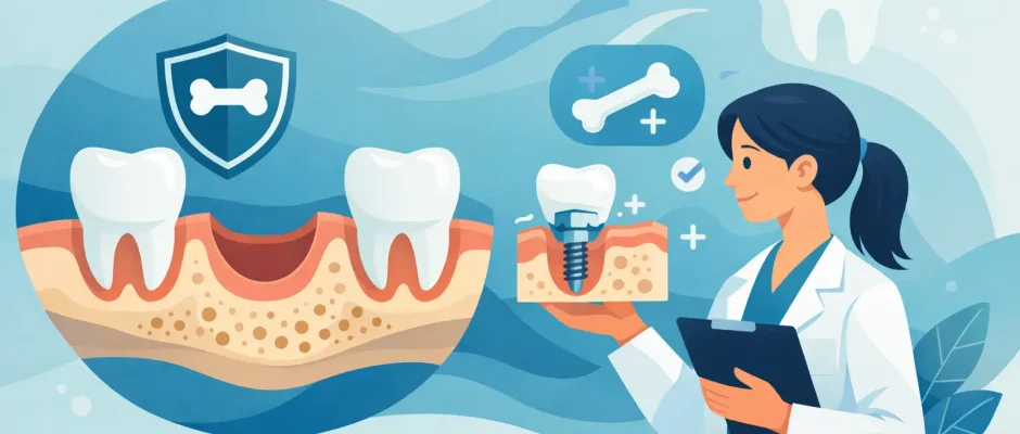
How to Prevent Bone Loss After Tooth Extraction
The extraction itself is usually not the difficult part. The decision made before the tooth is removed often determines how much bone will be available later for an implant, bridge, or natural-looking restoration. Knowing how to prevent bone loss after tooth extraction gives you time to choose a treatment plan that protects both function and facial esthetics.
A missing tooth is not simply an empty space in the mouth. The jawbone around each root depends on chewing forces to maintain its volume. Once a tooth is removed, that stimulation disappears. The body begins remodeling the extraction site, and the ridge can become narrower and lower within the first months. This is a normal biological process, but it can be managed with the right surgical protocol.
Why Bone Shrinks After a Tooth Is Removed
The bone surrounding a tooth is called alveolar bone. It develops and is maintained because the tooth root transmits pressure to the jaw during chewing. After extraction, the body no longer needs to maintain the same amount of bone in that exact location. Healing occurs, but healing does not always mean preservation of the original ridge shape.
The greatest dimensional changes commonly occur during the first three to six months after extraction. The outer wall of bone, especially in the front of the upper jaw, is often very thin. If it resorbs, the gum contour may flatten or recede. This matters most in the smile zone, where even a well-made crown can look too long or sit in an unnatural position if the underlying bone and soft tissue have collapsed.
Bone loss is not identical for every patient. The rate and extent depend on the thickness of the existing bone, the reason for extraction, periodontal infection, traumatic removal, smoking, uncontrolled diabetes, bite forces, and whether adjacent teeth are present. A tooth removed because of a vertical root fracture may leave a very different site from a tooth lost after years of severe gum disease.
How to Prevent Bone Loss After Tooth Extraction
The most effective strategy is to plan the extraction and replacement together. Waiting is sometimes appropriate, particularly when a large infection must resolve or general health requires stabilization. But when an implant is likely to be the final solution, the surgeon should evaluate the site before the tooth is removed, not months after bone volume has already changed.
Atraumatic extraction protects the starting point
Preserving bone begins with careful removal of the tooth. The objective is not simply to extract the root, but to minimize unnecessary trauma to the thin bony walls and surrounding gum tissue. In difficult cases, this may require sectioning the tooth, using microsurgical instruments, and removing roots in controlled stages rather than applying excessive force.
This is particularly relevant for teeth with curved roots, previous root canal treatment, large infections, or fragile bone. A precise extraction cannot eliminate normal remodeling, but it can preserve more of the anatomy needed for future treatment.
Socket preservation can limit ridge collapse
Socket preservation, also called ridge preservation, is a bone grafting procedure performed at the time of extraction. After the tooth is removed and the site is cleaned, a biocompatible graft material is placed in the socket. Depending on the clinical situation, it may be covered with a collagen membrane, a soft-tissue graft, or both.
The purpose is not to create an instant implant site. It is to support the healing space and reduce the degree of bone shrinkage that would otherwise occur. Over time, your own bone gradually remodels within and around the grafted area.
Socket preservation is often recommended when an implant will be placed later, when the outer bone wall is thin or damaged, when the tooth is in the esthetic zone, or when immediate implant placement is not advisable. It is also useful if you need time before deciding on a final restoration.
There is a trade-off: grafting adds a surgical step and cost, and it usually requires several months of healing before implant placement. However, it may reduce the need for more extensive bone augmentation later. In many cases, preserving the site at extraction is simpler and more predictable than reconstructing a severely resorbed ridge afterward.
Immediate implant placement may be an option
An immediate implant is placed into the extraction socket during the same surgical appointment. For the right patient, it can reduce treatment time and help preserve the existing tissue contours. It does not, however, automatically prevent bone loss. The implant must be positioned precisely, achieve primary stability, and be surrounded by adequate healthy bone.
Immediate placement is not appropriate for every extraction. Severe infection, insufficient bone for implant stability, missing socket walls, uncontrolled gum disease, or a high esthetic risk may make a staged approach safer. In some situations, extraction with socket preservation followed by delayed implantation produces the more reliable result.
Digital imaging and three-dimensional planning are valuable here. A CBCT scan allows the surgeon to assess root anatomy, bone thickness, hidden infection, the maxillary sinus in the upper jaw, and the position of nerves in the lower jaw. When indicated, a surgical guide can help transfer the approved implant plan accurately to the procedure.
PRF can support healing, but it is not a substitute for planning
Platelet-rich fibrin, or PRF, is made from a small sample of the patient’s own blood. It creates a fibrin matrix rich in platelets and growth factors and can be used within an extraction socket or alongside grafting procedures. PRF may support soft-tissue healing, reduce postoperative discomfort, and improve the handling of graft materials.
It is a useful biological adjunct, not a magic solution. PRF does not replace careful debridement of infection, stable grafting, membrane placement when needed, or correct implant positioning. Its value is greatest when it is integrated into a disciplined surgical protocol.
What You Can Do After Extraction
The first days after extraction matter. Protect the blood clot, because it is the foundation for normal healing. Follow the surgeon’s instructions regarding gauze, medication, oral hygiene, diet, and physical activity. Avoid smoking and vaping, which reduce blood supply and substantially increase the risk of impaired healing, infection, and dry socket.
Do not rinse forcefully, spit repeatedly, or use a straw during the early postoperative period if your surgeon advises against it. Choose soft foods, maintain hydration, and keep the rest of the mouth clean. If a temporary replacement is planned, it should be designed not to press heavily on a fresh grafted site.
Call the clinic if pain intensifies after initially improving, swelling worsens after several days, bleeding persists, or you develop fever, a foul taste, or numbness that does not resolve as expected. Early assessment can prevent a small postoperative issue from affecting the final result.
Do Not Leave the Space Unassessed
Not every missing tooth requires an implant immediately. A wisdom tooth, for example, often does not need replacement. In other locations, a bridge or removable prosthesis may be medically or financially appropriate. Yet leaving a functional tooth gap without a plan can lead to drifting of neighboring teeth, overeruption of the opposing tooth, bite changes, and progressive loss of bone volume.
If a tooth has already been removed months or years ago, treatment is still possible. A CBCT evaluation can show whether the existing ridge can support an implant or whether guided bone regeneration, a block graft, or a sinus lift is needed first. Modern reconstructive techniques can address significant bone deficiency, but early preservation generally gives more options and may shorten treatment.
The Questions That Shape a Safe Plan
Before an extraction, ask whether the tooth can be predictably saved and, if not, whether socket preservation or immediate implantation is appropriate. You should also understand the condition of the surrounding gums, the expected healing timeline, and what temporary tooth replacement will look like if the tooth is visible when you smile.
A good plan is individualized. It balances infection control, available bone, esthetic demands, medical history, bite forces, and your goals for timing and cost. For complex cases, the most valuable result is not the fastest extraction. It is a stable foundation for the tooth replacement you will rely on for years.
If extraction is unavoidable, schedule the consultation before the procedure whenever possible. Protecting the bone is usually easier at the moment the tooth is removed than trying to rebuild what has already been lost.


