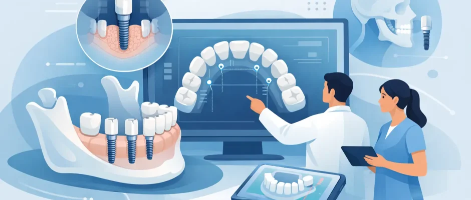
Digital Implant Planning for Safer Results
A dental implant is only a few millimeters wide, yet its position affects much more than the missing tooth. It must be placed within the available bone, at a safe distance from nerves and sinus cavities, and in the correct relationship to the future crown. Digital implant planning turns these requirements from estimates made during surgery into a carefully prepared treatment plan.
For a patient, the practical benefit is clarity. Before the procedure, the surgeon can assess the anatomy, explain whether bone grafting is needed, and determine whether immediate implant placement is realistic. For the clinical team, digital planning creates a more controlled protocol – particularly when bone volume is limited, several teeth are missing, or an implant is planned in the visible smile zone.
What digital implant planning actually involves
Digital implant planning combines three-dimensional imaging with a digital model of the teeth and gums. A cone beam CT scan, often called CBCT, shows the bone in detail: its height, width, density patterns, and relationship to important anatomical structures. An intraoral scan or precise dental impression records the surface of the teeth and soft tissue.
These datasets are aligned in planning software. The implant is then positioned virtually, not simply where bone appears available, but where it can support the planned restoration. Its diameter, length, depth, angle, and distance from adjacent roots are evaluated before treatment begins.
This distinction matters. An implant can integrate successfully with the bone and still be poorly positioned for a crown. If it is too far toward the lip, tongue, cheek, or palate, the final tooth may be difficult to clean, compromised esthetically, or subjected to unfavorable forces. The restorative result should guide the surgical plan whenever the case allows it.
Planning from the final tooth backward
A predictable protocol begins with the intended prosthetic outcome. The clinician considers the shape and position of the future tooth, the bite, neighboring teeth, gum line, and access for hygiene. The implant is planned to support that restoration while respecting the biological limits of the patient’s anatomy.
This is especially relevant for front teeth, where small deviations can influence gum symmetry and the emergence profile of the crown. It is equally relevant for full-arch rehabilitation, including All-on-4 concepts, where implant angulation and distribution affect the strength and design of the fixed prosthesis.
Why 3D planning improves safety
Traditional X-rays remain useful, but they are two-dimensional. They cannot always show the exact thickness of the jawbone or the spatial location of a nerve canal. CBCT-based digital implant planning provides information in all three dimensions, allowing the surgeon to identify areas that require special caution.
In the lower jaw, this often means mapping the mandibular nerve canal. In the upper posterior jaw, it means assessing the maxillary sinus and the remaining bone beneath it. The scan may also reveal an undercut, a thin bone wall, an impacted tooth, or inflammation near the intended implant site that changes the treatment sequence.
Planning does not eliminate surgical risk. Human anatomy varies, scans have limitations, and clinical decisions must still be made during treatment. However, knowing the anatomy in advance reduces avoidable surprises and supports a more measured, responsible approach.
When a surgical guide is useful
After virtual planning, a custom surgical guide can be designed and fabricated. This is a device that rests on teeth, gums, or bone and directs the initial drilling according to the planned implant position. Depending on the system and clinical indication, it may guide one step of the procedure or the full drilling sequence and implant placement.
A guide is not required for every implant. In a straightforward case with excellent visibility, sufficient bone, and a clearly defined surgical site, freehand placement may be entirely appropriate in experienced hands. In other situations, a guide can add meaningful control.
Guided surgery is often valuable when multiple implants must work together, when the available bone is narrow, when implants are near anatomical structures, or when immediate provisional teeth are planned. It can also make the procedure more efficient by reducing the need for intraoperative repositioning.
The guide itself is only as accurate as the diagnostic data, software design, fit, and surgical protocol. It must be checked carefully before use. A responsible surgeon does not follow a guide blindly; clinical judgment remains essential throughout the procedure.
Planning complex cases before choosing the procedure
Digital diagnostics are particularly valuable when an implant cannot simply be placed into an ideal volume of bone. Bone loss after tooth extraction is common, and it may be more pronounced after long periods without a tooth, chronic infection, gum disease, trauma, or previous surgery.
The scan helps answer practical questions: Is there enough bone for a stable implant? Can the implant be placed immediately after extraction? Is guided bone regeneration needed? Would a sinus lift create a safer foundation in the upper jaw? Should treatment be staged to allow healing before implant placement?
There is no universal answer. Immediate implant placement can shorten treatment time and preserve aspects of the existing anatomy in selected cases, but it requires adequate stability, infection control, appropriate soft tissue conditions, and a plan for the gap between implant and socket walls when necessary. If these conditions are not present, delayed placement may offer the more predictable option.
Similarly, bone grafting is not a sign that treatment has failed or become excessively complicated. It is often a planned method for creating the bone volume required for stable, correctly positioned implants. Materials, membranes, fixation methods, and healing periods are selected according to the defect rather than applied as a standard package.
Soft tissue is part of the plan
A well-positioned implant needs healthy, stable tissue around it. Digital planning shows bone anatomy, but the examination must also include gum thickness, the level of the gum line, smile visibility, periodontal health, and the condition of adjacent teeth.
In selected cases, soft tissue grafting or microsurgical techniques may be recommended to improve thickness, contour, or long-term hygiene around the implant. This is most relevant in esthetic areas and in patients with thin tissue biotypes. The objective is not merely to place an implant, but to create conditions that can remain stable and comfortable over time.
What happens at the consultation
A proper implant consultation should lead to a sequence of decisions, not a generic promise. The first step is a clinical examination: the surgeon evaluates the missing tooth area, bite, gums, neighboring teeth, oral hygiene, and medical history. If imaging is indicated, CBCT data are assessed together with the restorative requirements.
You should leave with an understandable plan: whether an extraction is needed, whether the implant can be placed on the same day, whether grafting is recommended, the likely healing stages, and what type of temporary or final restoration may be used. Timing depends on the clinical situation, not only on convenience.
For patients traveling for treatment in Tel Aviv, pre-planning is also useful for coordinating appointments and separating surgical and restorative phases realistically. Some cases can be completed efficiently; others require healing intervals that should not be rushed.
The patient’s role in a predictable result
Technology improves planning, but long-term implant success also depends on the patient. Smoking, uncontrolled diabetes, untreated gum disease, nighttime grinding, and inconsistent home care can affect healing and maintenance. These factors do not automatically rule out implant treatment, but they must be addressed honestly before surgery.
After placement, follow the prescribed medication and hygiene instructions, attend postoperative reviews, and avoid loading the area before it is cleared. Once the final crown is in place, regular professional maintenance remains part of implant care. An implant cannot develop a cavity, but the tissues around it can become inflamed if plaque control is inadequate.
Digital implant planning is valuable because it replaces assumptions with measurable information and gives both patient and surgeon a shared map before treatment begins. The right plan is not necessarily the fastest or most complex one. It is the plan that respects anatomy, supports the final restoration, and gives you a clear, calm understanding of each step ahead.


