What Causes Jaw Bone Loss? 8 Common Reasons
A missing tooth can begin changing the jaw long before it becomes visible in the mirror. The answer to what causes jaw bone loss is usually not one isolated event, but a biological response to lost stimulation, inflammation, or long-standing pressure on the bone. For patients considering implants, the key issue is timing: bone loss may complicate treatment, but it does not automatically rule it out.
The jawbone is living tissue. It constantly remodels itself in response to chewing forces, blood supply, and the health of the teeth and gums around it. When that balance changes, the bone can gradually lose height, width, or density. A careful clinical examination and 3D imaging show not only how much bone remains, but why it changed and which treatment plan can provide a predictable foundation.
What Causes Jaw Bone Loss After a Tooth Is Lost?
The most common cause is tooth loss itself. A natural tooth transfers chewing pressure through its root into the surrounding jawbone. This stimulation tells the body that the bone is needed. Once a tooth is extracted or lost, that signal disappears, and the body begins resorbing the portion of bone that once supported the root.
This process is often most active during the first months after extraction, although remodeling can continue for years. In the upper back jaw, bone changes may occur alongside expansion of the maxillary sinus. In the lower back jaw, the ridge may become narrower and lower over time. The result is less available bone for a conventionally placed implant.
A dental bridge can restore appearance and chewing function, but it does not replace the root inside the bone. A removable denture also cannot prevent ridge resorption on its own. An implant, when placed under suitable conditions, can provide functional stimulation to the bone, but it cannot fully restore bone that has already been lost without additional regenerative treatment.
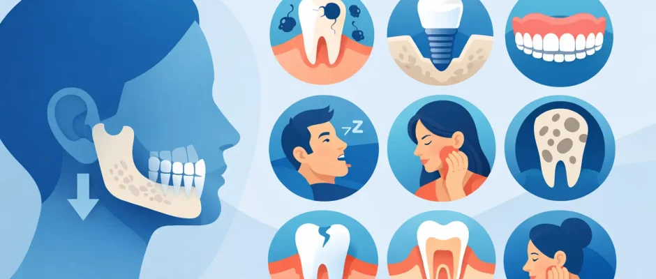
8 Common Reasons for Jawbone Loss
1. Delayed replacement after extraction
After an extraction, the socket heals, but healing does not mean that the original bone contour is preserved. The body replaces the extraction socket with bone, then gradually reshapes the ridge because there is no root in place. This is why socket preservation, immediate implant placement in selected cases, or timely implant planning may be discussed before or at the time of extraction.
Immediate implantation is not appropriate for every tooth. Active infection, insufficient bone walls, gum conditions, bite forces, and esthetic demands all affect the decision. The goal is not to place an implant quickly at any cost, but to choose the safest protocol for stable long-term results.
2. Advanced gum disease
Periodontitis is a chronic inflammatory disease caused by bacterial biofilm and an exaggerated inflammatory response. It destroys the ligament and bone that hold teeth in place. Early gum disease may cause bleeding and swelling, while advanced disease can lead to deep pockets, tooth mobility, bad breath, exposed roots, and progressive loss of jawbone around several teeth.
Periodontal bone loss is particularly important before implant treatment. Implants do not develop cavities, but the surrounding tissues can become inflamed and lose bone if plaque control and maintenance are inadequate. Treating active periodontal disease and establishing reliable hygiene are essential parts of implant planning.
3. Infection at the root of a tooth
A deep cavity, crack, failed root canal treatment, or severe trauma can allow infection to reach the tip of a tooth root. The body responds with inflammation that may gradually dissolve the surrounding bone. Sometimes this appears as a cyst-like lesion or a dark area on an X-ray; sometimes symptoms are minimal until the infection becomes acute.
When possible, root canal retreatment, microsurgery such as apicoectomy, or other tooth-preserving procedures may save the tooth. If extraction is necessary, the infected tissue must be thoroughly removed and the site assessed before implant placement. The timing of reconstruction depends on the extent of infection and the quality of the remaining bone and soft tissue.
4. Poorly fitting dentures and chronic pressure
A removable denture rests on the gum-covered ridge. If it is loose, unstable, or worn for years without adjustment, repeated pressure can accelerate resorption in certain areas. Patients may notice that the denture moves while speaking or eating, requires more adhesive, or causes sore spots.
This does not mean dentures are inherently unsafe. A well-designed denture with regular follow-up can be a reasonable solution for some patients. However, in cases of progressive ridge loss, implant-supported options can improve stability and reduce harmful movement. For a full arch, the appropriate approach may range from a few supporting implants to a fixed full-arch protocol, depending on anatomy and health.
5. Trauma and fractures
Facial trauma can damage the jawbone directly or disrupt the blood supply to teeth. A tooth may initially appear intact after an injury but later develop root resorption, nerve damage, infection, or bone loss around the root. Previous jaw fractures, surgical plates, scars, and altered anatomy can also influence implant planning.
These situations require precise imaging. A CBCT scan helps evaluate bone volume, the position of nerves and sinuses, root fractures, and the relationship between a future implant and neighboring structures. Digital planning and a surgical guide can add control when anatomy is limited or complex.
6. Peri-implant disease
Bone loss can also occur around an existing implant. Mucositis is inflammation of the soft tissue around an implant and can often be managed before bone is affected. Peri-implantitis involves inflammation with progressive bone loss and requires timely treatment.
Risk factors include a history of periodontitis, smoking, uncontrolled diabetes, poor access for cleaning, residual cement around a restoration, excessive bite forces, and missed maintenance visits. The exact treatment depends on the defect, implant position, surface condition, and ability to control infection. Some implants can be treated and maintained; others may need to be removed so the site can heal and be reconstructed.
7. Smoking and systemic health conditions
Smoking reduces blood flow, impairs healing, and increases the risk of periodontal disease and complications after surgery. Heavy smoking does not make implant treatment impossible in every case, but it changes the risk profile and should be discussed openly before bone grafting or implant placement.
Systemic conditions matter as well. Poorly controlled diabetes can impair healing and increase infection risk. Osteoporosis affects skeletal metabolism, although it does not automatically prevent implant treatment. Certain medications, especially antiresorptive drugs used for osteoporosis or cancer-related bone disease, require careful medical review because they may affect jaw healing after invasive procedures. Treatment should be coordinated with the patient’s physician when indicated.
8. Long-term overload and unfavorable bite forces
Grinding, clenching, missing opposing teeth, or an uneven bite can overload teeth, implants, and restorations. Overload alone is rarely the only reason for bone loss, but when combined with inflammation or poor implant positioning, it can contribute to complications. A strong final restoration requires more than an implant integrating with bone. It must also fit the patient’s bite, chewing pattern, and hygiene needs.
How Jaw Bone Loss Changes Implant Treatment
Bone loss does not have a single solution. A small localized defect may be managed with grafting at the time of implant placement. A wider horizontal deficiency may need guided bone regeneration using graft material and a protective membrane. In the upper posterior jaw, a sinus lift may create sufficient vertical space for an implant. In more extensive cases, treatment may be staged to allow regenerated bone to mature before implants are placed.
PRF, made from the patient’s own blood, may be used as an adjunct to support soft-tissue and bone healing in appropriate surgical protocols. It is not a substitute for sound surgical planning, infection control, or a well-designed reconstruction. The predictable result comes from matching the technique to the defect, rather than applying one method to every patient.
For some patients with severe full-arch bone loss, angled implants and fixed full-arch concepts such as All-on-4 may avoid certain grafting procedures. That can shorten treatment in carefully selected cases, but it is not automatically better than rebuilding bone. The decision depends on the available anatomy, smile line, hygiene ability, medical history, expectations, and the desired long-term restoration.
When to Seek an Evaluation
Do not wait for pain to assess a missing tooth or unstable implant. Gum bleeding, loosening teeth, recurrent swelling, a denture that no longer fits, visible recession, or difficulty chewing all justify an examination. A clinical evaluation with periodontal measurements and CBCT imaging, when indicated, can identify whether bone loss is active, stable, localized, or widespread.
At Implantolog.co.il, treatment planning is built around this diagnosis: preserving a tooth when that offers a reliable prognosis, reconstructing bone when it improves implant positioning, and using digital planning to reduce uncertainty during surgery. The right plan should explain the sequence, healing periods, alternatives, and realistic limits before treatment begins.
Jawbone loss is often gradual, which makes early action valuable. A precise diagnosis now can preserve more options, reduce the scale of future surgery, and make the path back to comfortable chewing feel far more manageable.


