A missing tooth is not always the main obstacle to an implant. Sometimes the limiting factor is the bone that once supported it. When the jaw ridge has become too narrow or too low, an implant cannot be placed in a stable, prosthetically correct position. In selected cases, an autogenous bone block graft provides the volume needed to rebuild that foundation using the patient’s own bone.
This is a more involved procedure than minor bone augmentation. It requires careful diagnosis, precise surgical technique, and patience during healing. Its purpose is not simply to “add bone,” but to create a stable ridge that can support a long-term implant restoration with appropriate contour, function, and hygiene access.
What is an autogenous bone block graft?
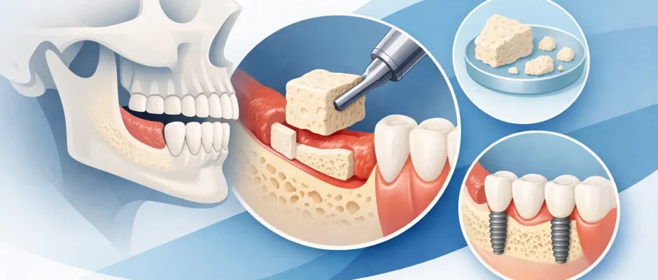
An autogenous bone block graft is a piece of bone taken from one area of a patient’s body and secured to an area of the jaw where bone volume is deficient. In implant surgery, the donor bone is most commonly harvested from inside the mouth, often from the lower jaw near the chin or the posterior mandibular region. Because the tissue comes from the same patient, it is living autogenous material rather than a substitute of animal or synthetic origin.
The surgeon shapes the bone block to fit the recipient site and fixes it with small titanium screws. Small gaps around the block may be filled with particulate bone material, depending on the anatomy. A barrier membrane may also be used to protect the grafted area and support guided bone regeneration. The soft tissues are then closed without tension, which is a critical part of predictable healing.
A block graft is generally considered when the defect is too large or too three-dimensional for a particulate graft alone. It can restore horizontal width, vertical height in selected situations, or both. The final decision is based on a cone beam CT scan, the planned implant position, bite forces, gum thickness, and the condition of neighboring teeth.
When is a bone block graft recommended?
The procedure is not the standard answer to every case of bone loss. Many patients can be treated with less invasive approaches, including guided bone regeneration with particulate graft material, a sinus lift in the posterior upper jaw, short implants, or a modified implant position. However, these options are not interchangeable.
An autogenous bone block graft may be appropriate when a patient has a pronounced ridge defect after a long-standing extraction, trauma, infection, previous surgery, or severe periodontal bone loss. It can be especially valuable in the visible front region, where both implant stability and the contour of the gum line influence the final esthetic result.
It may also be considered after failed implant treatment or when earlier augmentation has not produced enough stable bone. In these cases, the goal is to avoid placing an implant in a compromised position merely because there is some bone available. A poorly positioned implant can create long-term difficulties with the crown, gum recession, cleaning, and load distribution.
For full-arch rehabilitation, including certain All-on-4 cases, a block graft is not always required. Strategic implant angulation and available basal bone can sometimes avoid extensive augmentation. Yet when the desired implant position cannot be achieved safely, rebuilding the ridge may be the more responsible choice.
Why use the patient’s own bone?
Autogenous bone has biological advantages. It contains the patient’s own mineralized matrix and cells associated with bone healing, and it integrates directly with the recipient site. For larger defects, its structural rigidity can help maintain the shape of the reconstructed ridge more effectively than loose graft material alone.
That said, using a patient’s own bone also creates a second surgical site. There may be postoperative discomfort, swelling, temporary altered sensation, or other donor-site risks. The lower jaw contains important nerves and tooth roots, so harvesting must be planned from three-dimensional imaging and performed with a disciplined microsurgical approach.
The choice is therefore a balance. A bone block can provide substantial regenerative potential and structural support, but it is more invasive than simpler augmentation. The best method is not the one that sounds most advanced. It is the method that achieves the required bone volume with the lowest reasonable surgical burden and a predictable prosthetic outcome.
The role of digital planning
The graft should be planned around the future tooth, not around the defect alone. A digital implant plan helps determine where the implant must emerge through the gum and where its crown will sit in relation to the bite and adjacent teeth. This prevents a common error: rebuilding bone in a way that still does not allow correct implant placement.
At the consultation, the surgeon evaluates CBCT imaging, oral hygiene, gum health, medical history, smoking status, and the condition of the opposing teeth. If an implant is planned after graft healing, its ideal dimensions and direction are considered before surgery. In appropriate cases, a surgical guide supports accurate implant placement at the next stage.
How the procedure and healing are usually staged
The surgery is performed under local anesthesia, with additional sedation options when clinically appropriate and desired. After preparing the recipient site, the surgeon harvests and adapts the bone block, secures it with fixation screws, and closes the area carefully. PRF, prepared from the patient’s own blood, may be used as an adjunct to support soft-tissue healing and graft management. It is not a substitute for stable fixation or meticulous surgical technique.
Swelling and discomfort are expected during the first days. Most patients manage this period with prescribed or recommended medication, cold packs during the initial phase, soft food, and clear hygiene instructions. Physical exertion, smoking, and pressure on the grafted area can compromise healing and should be avoided according to the surgeon’s instructions.
The graft typically needs several months to integrate before implant placement. The exact interval depends on the size and location of the graft, the quality of the native bone, the type of augmentation, and the patient’s healing response. Follow-up visits are not a formality. They allow the surgeon to monitor soft-tissue closure, identify early inflammation, and confirm that healing is progressing as planned.
At the implant stage, fixation screws are removed if necessary, and the regenerated site is assessed. In selected cases, a minor additional contour graft may be needed around the implant. After osseointegration, the restorative dentist completes the crown, bridge, or full-arch prosthesis. This staged approach takes time, but it protects the biological foundation of the final work.
Risks and factors that affect predictability
Bone grafting is highly technique-sensitive. Even with appropriate planning, potential complications include wound opening, infection, partial graft resorption, graft exposure, failure of integration, or temporary sensory changes at the donor site. These risks are discussed before treatment because informed consent should include both the expected benefits and the limits of the procedure.
Smoking significantly increases the risk of impaired healing and complications. Active periodontal disease, uncontrolled diabetes, poor oral hygiene, untreated bite overload, and inconsistent follow-up can also affect results. A prior history of failed grafting does not automatically rule out another attempt, but it requires a careful analysis of why the earlier treatment failed.
The quality of the soft tissue is just as relevant as the amount of bone. Thick, well-managed gum tissue helps protect the graft and supports a more stable esthetic result around an implant. For this reason, hard-tissue and soft-tissue surgery are often planned together rather than treated as separate problems.
What patients should ask before treatment
A clear treatment plan should explain where the bone will be taken from, why a block graft is preferable to less invasive alternatives, when the implant can be placed, and what temporary tooth replacement is possible during healing. Ask how the implant position will be planned, what follow-up is included, and what signs require an urgent call after surgery.
It is also reasonable to ask about the expected number of surgical stages. In complex reconstruction, transparency matters more than promising a fast result. A shorter treatment timeline is valuable only when it does not compromise bone stability, implant position, or the long-term health of the surrounding tissues.
For patients considering complex implant treatment in Tel Aviv, the most useful first step is a focused consultation with clinical examination and CBCT-based planning. The right plan may involve an autogenous bone block graft, a simpler regenerative procedure, or no graft at all. The aim is a stable implant foundation that allows you to eat, smile, and maintain your restoration with confidence for years to come.


