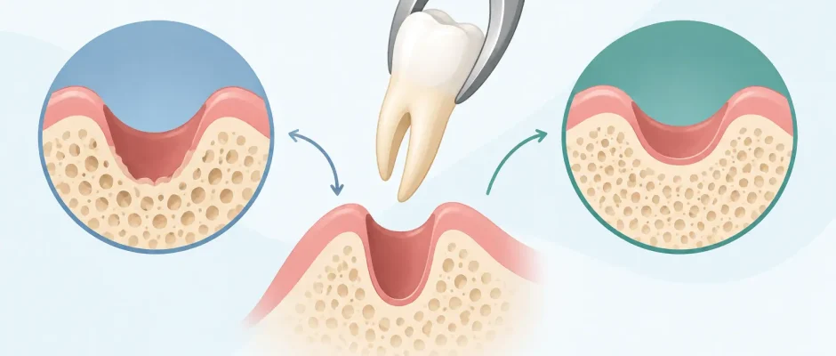
How Atraumatic Extraction Reduces Bone Loss
A tooth extraction is not only about removing a tooth. It is also about protecting the bone and soft tissue that remain after it. Understanding how atraumatic extraction reduces bone loss helps patients see why a careful surgical approach matters, especially when an implant may be planned later.
After a tooth is removed, the jawbone in that area naturally begins to remodel. Some of this change is unavoidable because the bone no longer receives stimulation from the tooth root. However, unnecessary trauma to the socket walls, gums, and surrounding bone can increase the loss of volume and make future implant treatment more complex. The goal of atraumatic extraction is to remove the tooth as gently and precisely as the clinical situation allows while preserving the tissues needed for reconstruction.
Why bone changes after a tooth extraction
The bone surrounding a tooth is called alveolar bone. It develops and is maintained largely because a tooth root is present. Once the root is removed, the body begins a natural healing and remodeling process. During the first months after extraction, the ridge may become narrower and lower, with the outer facial wall often being particularly vulnerable.
This does not mean that every extraction leads to a major defect. The amount of change depends on the tooth position, thickness of the bone walls, periodontal condition, infection, previous bone loss, smoking status, general health, and the way the tooth is removed. A front tooth with a thin facial bone plate presents different risks than a molar with strong surrounding bone. An impacted wisdom tooth, a fractured root, or a severely infected tooth may require a more complex surgical approach.
The important distinction is between biologic remodeling, which cannot be eliminated completely, and additional damage caused by excessive force or uncontrolled manipulation. Atraumatic extraction aims to avoid the second problem.
How atraumatic extraction reduces bone loss
Atraumatic extraction is a protocol, not simply a promise to “pull gently.” It begins with diagnosis. Clinical examination and imaging, often including CBCT when indicated, allow the surgeon to assess root shape, bone thickness, proximity to anatomical structures, infection, and the possibility of preserving the socket for an implant.
During the procedure, the periodontal ligament around the root is carefully released. Fine instruments may be used to expand the ligament space gradually rather than applying strong force to the surrounding bone. When a tooth has multiple roots, curved roots, a large restoration, or a fragile crown, it may be safer to section the tooth into smaller parts and remove each part individually.
This approach helps preserve the bony walls of the extraction socket. In particular, avoiding a fracture of the thin facial plate can be significant for both function and appearance. Preserved bone provides a more favorable foundation for an implant, a bridge, or a removable restoration, and it may reduce the extent of future bone grafting.
Atraumatic technique also protects the soft tissue envelope. The gum contours around a tooth contribute to the natural appearance of an implant restoration, especially in the smile zone. If soft tissue is torn, over-stretched, or poorly supported during healing, achieving an ideal esthetic contour later may be more difficult.
Precision matters more than speed
A difficult extraction should not be forced to become a quick extraction. When the crown breaks, roots are thin or fused, or the tooth is tightly anchored in dense bone, controlled surgical access is often safer than repeated attempts with forceps.
For example, a molar may be divided at the furcation so that each root can be removed in a controlled direction. A retained root fragment may require careful visibility and selective bone removal rather than blind instrumentation. In cases involving a lower wisdom tooth close to the mandibular nerve, preoperative planning is essential to reduce unnecessary risk.
The procedure may take longer than a conventional extraction, but the purpose is not to prolong treatment. It is to protect important anatomy and create conditions for predictable healing. Comfort is also part of that protocol: profound local anesthesia, clear communication, gentle tissue handling, and a defined postoperative plan reduce the stress of surgery for many patients.
Atraumatic does not mean bone loss is impossible
Even a technically excellent extraction cannot stop all post-extraction remodeling. The body will still adapt after a tooth is lost. For this reason, preserving the socket may involve more than careful removal alone.
Socket preservation, also called ridge preservation, may be recommended when an implant is planned later or when the existing bone walls are thin. After the tooth is removed and the socket is cleaned, a bone graft material may be placed to support the space during healing. Depending on the case, the graft can be combined with a membrane and sutures to stabilize the site and guide tissue regeneration.
PRF, prepared from the patient’s own blood, can also be used in selected cases. It forms a fibrin matrix containing platelets and growth factors that may support soft-tissue healing and help stabilize grafting material. PRF is not a substitute for sound surgical technique or an indication for grafting in every extraction. It is one component of a treatment plan chosen according to the anatomy and clinical goal.
If the socket walls are already damaged by infection, periodontal disease, cystic changes, or a previous failed extraction, more extensive regeneration may be needed. This is precisely why a surgeon should evaluate the site before deciding whether immediate implantation, socket preservation, delayed implantation, or staged bone augmentation is the most responsible option.
Immediate implant placement: useful in the right case
Many patients ask whether an implant can be placed on the same day as extraction. Immediate implant placement can shorten treatment time and may help maintain tissue contours in properly selected cases. But it is not automatically the best solution simply because it is possible.
For an immediate implant to be predictable, the surgeon must be able to obtain stable implant fixation, control infection, assess the integrity of the socket walls, and position the implant prosthetically and anatomically correctly. The implant should not be used as a way to compensate for a rushed extraction or unrecognized bone defect.
In some cases, immediate placement is an excellent option. In others, preserving the socket, allowing healing, and placing the implant later produces a safer and more stable result. A staged plan is not a setback. It may be the correct way to protect bone and reduce the risk of complications.
What happens after the extraction
The healing period matters as much as the procedure itself. A stable blood clot is the first stage of normal socket healing. Patients are generally advised to avoid vigorous rinsing, spitting, smoking, and intense physical activity during the early postoperative period, because these actions can interfere with the clot and increase the risk of complications.
Mild swelling, tenderness, and limited mouth opening can occur, particularly after surgical extractions. The expected recovery depends on the tooth, the complexity of the procedure, whether grafting was performed, and the patient’s overall health. A personalized postoperative plan should include pain control, hygiene instructions, dietary guidance, and a scheduled review when necessary.
Contact the clinic promptly if pain becomes stronger after several days rather than improving, if there is persistent bleeding, fever, worsening swelling, numbness, or an unpleasant taste and odor that does not resolve. Early assessment can prevent a small issue from becoming a larger one.
When bone preservation deserves special attention
Bone-preserving extraction is particularly relevant when a visible front tooth is being removed, when an implant is planned, or when the patient already has reduced ridge volume. It is also valuable in patients who may need full-arch rehabilitation, including All-on-4 protocols, because every available millimeter of bone can influence implant positioning and the need for additional grafting.
It also matters when no immediate replacement is planned. Delaying the decision to replace a tooth does not stop bone remodeling. If preserving future treatment options is important, discussing socket preservation before extraction is often wiser than trying to rebuild a significantly resorbed ridge years later.
At Implantolog.co.il, extraction planning is approached as part of the larger surgical and restorative plan. The question is not only how to remove a problematic tooth, but how to protect the conditions needed for the next stage of treatment.
A careful extraction cannot preserve every particle of bone, and no ethical clinician should promise that it will. What it can do is reduce avoidable injury, support more favorable healing, and keep more treatment options open. Before an extraction, ask what the bone looks like now, whether the socket walls are intact, and what plan best protects your future smile.


