Microsurgical Tooth Extraction Techniques Explained
A tooth that cannot be saved should not be removed by force. The quality of extraction affects the bone and gum tissue that remain, the comfort of healing, and the options available for future restoration. Microsurgical tooth extraction techniques are designed to make this stage as controlled and tissue-preserving as possible, particularly when an implant may be considered later.
For patients, the difference is often felt as a calmer procedure with a clear plan. For the surgeon, it means working under magnification, using delicate instruments, and making decisions based on three-dimensional diagnostics rather than treating every extraction as routine.
What Makes an Extraction Microsurgical?
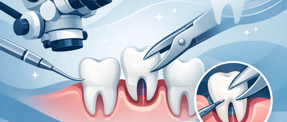
Microsurgical extraction is not simply a more careful version of standard tooth removal. It is a protocol focused on minimizing unnecessary trauma to the socket, surrounding bone, periodontal ligament, and gingiva. Magnification, focused illumination, fine instruments, and atraumatic techniques allow the surgeon to see and control details that are difficult to manage with conventional force alone.
The objective is not to make every tooth come out in one piece. In some cases, preserving the tooth structure is less important than preserving the bone around it. A severely damaged molar, a tooth with curved roots, or a retained root fragment may need to be sectioned into smaller parts and removed separately. This can be more precise and safer than applying increasing pressure to the jawbone.
The approach is particularly relevant in the esthetic zone, where even a small loss of facial bone or gum contour can influence the appearance of a future crown. It is also valuable for teeth near the maxillary sinus, the mandibular nerve canal, or adjacent healthy teeth.
Microsurgical Tooth Extraction Techniques in Practice
Every procedure begins with diagnosis. A clinical examination and digital imaging help determine the root shape, bone level, active infection, proximity to anatomical structures, and prognosis of the tooth. For complex cases, cone beam computed tomography, or CBCT, provides a three-dimensional view that cannot be obtained from a standard two-dimensional X-ray alone.
Gentle separation of the periodontal ligament
A tooth is held in its socket by the periodontal ligament. Instead of immediately using broad elevators and forceful movements, the surgeon may first carefully separate these fibers with thin periotomes or luxators. This creates controlled mobility while reducing pressure on the thin socket walls.
This step matters most where the outer bone plate is delicate. The facial bone around upper front teeth, for example, may be very thin. If it fractures during extraction, the site may require more extensive grafting before or during implant treatment.
Root sectioning when it protects bone
Multi-rooted teeth are often better removed in sections. A lower molar may be divided so that each root can be elevated along its individual path. An upper molar may require a similarly measured approach because of its relationship to the sinus and the shape of its roots.
Patients sometimes worry when they hear that a tooth will be sectioned. Clinically, this is frequently the more conservative option. Reducing the size of each fragment can reduce the force needed for removal and lower the risk of damaging adjacent structures.
Controlled management of difficult roots
Curved, fragile, or previously treated roots can fracture during extraction even with excellent technique. A fractured root is not automatically a complication, but it does require a deliberate decision. The surgeon evaluates its location, signs of infection, depth, and the risks of retrieval.
In many cases, the fragment can be removed through careful access under magnification. In selected situations, attempting aggressive retrieval may create more harm than benefit, especially if the fragment is close to the nerve canal or sinus. The correct approach depends on the individual anatomy, not on a one-size-fits-all rule.
Protection of soft tissue and socket walls
Microsurgical work includes respect for the surrounding gingiva. When an incision or small flap is necessary, it is designed to provide access while preserving blood supply and supporting accurate closure. Fine sutures may be used to stabilize the tissue and promote undisturbed healing.
After the tooth is removed, the socket is inspected. Granulation tissue or infected material is carefully debrided when present, while healthy bone and soft tissue are preserved. The goal is a clean, stable site ready to heal or to receive the next indicated step of treatment.
When Bone Preservation Becomes Part of the Plan
Extraction is often the first stage of implant rehabilitation, but immediate implant placement is not appropriate in every case. The decision depends on bone volume, gum thickness, socket integrity, infection control, bite forces, and the ability to achieve stable implant fixation.
If conditions are favorable, an implant may sometimes be placed at the time of extraction. This can shorten the treatment sequence, but it still requires precise planning. An immediate implant is not simply placed into the empty socket. It must be positioned in the ideal restorative location, with adequate primary stability and thoughtful management of any gaps between the implant and socket wall.
When immediate placement is not predictable, socket preservation may be recommended. This may involve bone graft material, a collagen membrane, and, in selected cases, platelet-rich fibrin, or PRF, prepared from the patient’s own blood. These measures do not eliminate natural bone remodeling, but they can help maintain the volume needed for later restoration.
For an infected tooth, the presence of infection does not automatically rule out immediate implantation. However, it raises the standard for debridement, site assessment, and case selection. If predictable stability or complete cleaning cannot be achieved, staged treatment is often the safer choice.
Which Cases Benefit Most?
Microsurgical tooth extraction techniques are valuable for many routine extractions, but they are especially useful when the consequences of tissue loss are significant. This includes upper front teeth planned for implants, teeth with extensive root decay, teeth fractured below the gum line, and teeth with large restorations that leave little structure to grip.
They are also relevant for impacted or displaced wisdom teeth, retained roots, teeth close to the sinus or inferior alveolar nerve, and teeth in patients with reduced bone volume. In these situations, meticulous planning is not an added luxury. It is part of reducing avoidable surgical risk.
A patient with advanced periodontal disease may need a different strategy from a patient whose tooth was damaged by trauma. Likewise, someone planning a single implant has different priorities from a patient preparing for full-arch rehabilitation. The extraction plan should support the final restorative goal from the beginning.
What the Patient Can Expect
Before surgery, the treatment plan should explain what will be removed, whether grafting or immediate implantation is being considered, and what healing timeline is realistic. Local anesthesia is used to ensure the procedure is comfortable. For patients with significant anxiety or a long, complex treatment plan, additional sedation options may be discussed where appropriate.
During the first days after extraction, mild swelling, soreness, and limited mouth opening can occur, particularly after surgical removal of wisdom teeth or deeply positioned roots. These effects are usually temporary. Following postoperative instructions is essential: protect the blood clot, take prescribed medication exactly as directed, maintain careful oral hygiene, and attend follow-up visits.
Pain that intensifies after initial improvement, persistent bleeding, fever, worsening swelling, numbness, or an unpleasant taste with increasing discomfort should prompt contact with the treating clinic. Early assessment helps distinguish normal healing from conditions that need attention.
Precision Is a Form of Patient Care
An extraction may take only a short time, but its biological consequences continue for months. Preserving bone, maintaining healthy soft tissue, and planning for the final restoration can reduce compromises later in treatment.
At Implantolog.co.il in Tel Aviv, complex extraction planning is approached as part of the larger surgical and implant treatment pathway, not as an isolated procedure. The most reassuring plan is one that explains not only how a tooth will be removed, but also how the site will be protected and what comes next.


