Socket Preservation After Extraction: When It Helps
A tooth extraction is not only the removal of a tooth. Once the root is gone, the jawbone that supported it begins to remodel. For a patient considering an implant later, socket preservation after extraction may be the step that keeps the treatment plan simpler, safer, and more predictable.
The procedure is often described as “putting bone graft material in the socket,” but that phrase leaves out the clinical judgment involved. The condition of the socket walls, the presence of infection, the thickness of the surrounding gum tissue, the position of the future implant, and the patient’s overall health all affect the decision. Preservation is valuable in many cases, but it is not automatic and it does not replace careful planning.
What socket preservation after extraction does
After extraction, the body heals the site by forming a blood clot, then soft tissue and new bone. At the same time, however, the ridge changes shape. The outer bone wall, especially in the front of the upper jaw, can be very thin and is prone to resorption. Over the first months, both the width and height of the bone may decrease.
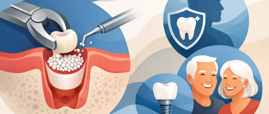
Socket preservation, also called ridge preservation, aims to reduce this loss. After the tooth is carefully removed, graft material is placed inside the socket. Depending on the case, it may be covered with a collagen membrane and secured with sutures. The graft acts as a scaffold while the body gradually replaces or integrates it with newly formed bone.
The goal is not to freeze the anatomy exactly as it was. Some remodeling is biologically normal and should be expected. The goal is to maintain enough bone volume and favorable soft-tissue contours for a future implant, bridge, or esthetic restoration, while reducing the chance that a more extensive bone graft will be needed later.
Why bone loss matters for an implant
An implant needs bone around it in all three dimensions. The surgeon must be able to position it according to the future crown, not simply place it where bone happens to remain. This is particularly relevant in the visible smile zone, where even a small loss of facial bone or gum contour can affect the final esthetic result.
When a socket heals without preservation, implant placement may still be entirely possible. Many patients do not need additional grafting. But if the ridge becomes narrow or collapses toward the tongue or palate, treatment may require guided bone regeneration, a block graft, or a different implant position. These procedures are effective when indicated, yet they can add healing time, cost, and surgical complexity.
Preserving the socket at the time of extraction is therefore often a preventive decision. It is most useful when the tooth cannot be saved and an implant is a realistic future option. It can also help preserve the contour beneath a pontic in a bridge or maintain tissue support in an esthetically demanding area.
When is preservation recommended?
There is no one protocol for every extraction. The indication is based on clinical examination and three-dimensional imaging when needed. A CBCT scan can show the condition of the socket walls, the relationship to adjacent roots and anatomical structures, and the available bone for implant planning.
Preservation is commonly considered when an implant is planned but cannot be placed immediately. This may happen because of active infection, a large bone defect, insufficient primary stability, unfavorable soft tissue, or a need to stage treatment for a more predictable result.
It can also be appropriate after removal of a front tooth, after extraction of a premolar where ridge width is already limited, or when the outer socket wall is damaged during infection or fracture. In the posterior upper jaw, preserving bone may simplify later implant planning near the maxillary sinus. In the lower jaw, it may help maintain ridge form where future implant placement would otherwise be more challenging.
On the other hand, socket preservation is not always necessary. If immediate implant placement is safe and stable, the implant procedure itself may include grafting of a small gap around the implant when indicated. If no implant or fixed restoration is planned, the value of grafting depends on the patient’s goals and the anticipated effect of ridge changes. A detailed discussion should address the benefit in that individual case, rather than treating grafting as a routine add-on.
What happens during the procedure
Atraumatic extraction is the first part of preservation. The aim is to remove the tooth while protecting the surrounding bone and gum tissue as much as possible. This may require sectioning a multi-rooted tooth or using microsurgical instruments rather than applying excessive force.
After the tooth is removed, the socket is cleaned carefully. Inflamed tissue, granulation tissue, and any residual infected material are removed. The surgeon then evaluates the bony walls. A contained socket with intact walls is managed differently from a site where the facial wall is missing or substantially damaged.
The graft material may be mineralized, demineralized, synthetic, or of animal origin. The choice depends on the defect, the desired healing behavior, and the planned timing of implant placement. A collagen membrane may be used to stabilize the material and guide healing. In selected cases, platelet-rich fibrin, or PRF, can support soft-tissue healing and improve management of the surgical site.
The area is usually closed with sutures. Complete tight closure is not required in every technique, but stable protection of the graft is essential. The procedure is performed under local anesthesia, and sedation can be discussed for patients with significant dental anxiety or complex surgical needs.
Healing time and what to expect
Most patients return to normal light activity within a day or two. Swelling, tenderness, and minor bruising can occur, especially if the extraction was difficult or the site had active inflammation. These symptoms are usually managed with prescribed or recommended pain control, cold compresses during the first day, and clear postoperative instructions.
The blood clot and grafted site must be protected. Smoking, forceful rinsing, drinking through a straw, and vigorous exercise in the first days can interfere with healing. Good oral hygiene remains important, but the surgical area should be cleaned only as instructed. Sutures are typically checked or removed during a postoperative visit.
The grafted socket commonly requires several months of healing before implant placement. The exact interval may range from approximately three to six months, depending on the graft material, the size of the defect, soft-tissue healing, and the planned implant protocol. Follow-up examination and imaging determine whether the site is ready. The calendar alone should not decide the timing.
Can an infected tooth still be grafted?
Often, yes, but only after proper debridement and assessment. Infection does not automatically rule out socket preservation or even immediate implant placement. The key issue is whether the source of infection can be thoroughly removed and whether the remaining bone and soft tissues provide conditions for stable healing.
In some cases, staging is the safer choice: extract the tooth, clean the site, preserve the ridge, allow healing, and place the implant later. This approach can be more predictable than trying to complete every stage at once. Patients sometimes view a staged plan as a delay, but it may be the most efficient path to a stable long-term result.
Questions worth asking before extraction
Before a tooth is removed, the future restoration should be discussed whenever possible. Ask whether the tooth has been assessed for saving with endodontic treatment, retreatment, microsurgery, or periodontal therapy. If extraction is necessary, ask whether an implant is planned, whether immediate placement is possible, and whether the socket walls are intact.
It is also reasonable to ask what graft material and membrane are being recommended, whether PRF may be useful, how long healing is expected to take, and what the backup plan is if the bone does not mature as anticipated. Clear answers reduce uncertainty and allow treatment to be planned around work, travel, and health needs.
For patients seeking implant treatment in Tel Aviv, a surgical consultation should begin with diagnosis rather than a preselected procedure. At Implantolog.co.il, the treatment plan is based on the tooth’s prognosis, three-dimensional anatomy, soft-tissue conditions, and the desired final restoration.
A well-preserved socket does not guarantee an implant without further grafting, and an ungrafted socket does not mean an implant is impossible. What it can do is protect options at the moment they matter most: before the natural bone contour has had time to change.


